 Movie
Movie Controller

[English] 日本語
 Yorodumi
- PDB-1fxy: COAGULATION FACTOR XA-TRYPSIN CHIMERA INHIBITED WITH D-PHE-PRO-AR... -
+ Open data

- Basic information
| Entry | Database: PDB / ID: 1fxy | ||||||
|---|---|---|---|---|---|---|---|
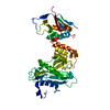
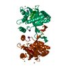
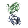
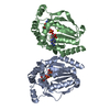
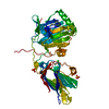
| Title | COAGULATION FACTOR XA-TRYPSIN CHIMERA INHIBITED WITH D-PHE-PRO-ARG-CHLOROMETHYLKETONE | ||||||
 Components Components | COAGULATION FACTOR XA-TRYPSIN CHIMERA | ||||||
 Keywords Keywords | hydrolase/hydrolase inhibitor / CHIMERA / PROTEASE / CHLOROMETHYLKETONE / HYDROLASE-HYDROLASE INHIBITOR COMPLEX | ||||||
| Function / homology |  Function and homology information Function and homology informationUptake of dietary cobalamins into enterocytes / Developmental Lineage of Pancreatic Acinar Cells / Activation of Matrix Metalloproteinases / extracellular matrix disassembly / trypsin / digestion / extracellular matrix / blood microparticle / serine-type endopeptidase activity / proteolysis ...Uptake of dietary cobalamins into enterocytes / Developmental Lineage of Pancreatic Acinar Cells / Activation of Matrix Metalloproteinases / extracellular matrix disassembly / trypsin / digestion / extracellular matrix / blood microparticle / serine-type endopeptidase activity / proteolysis / : / extracellular region / metal ion binding Similarity search - Function | ||||||
| Biological species |  Homo sapiens (human) Homo sapiens (human) | ||||||
| Method |  X-RAY DIFFRACTION / Resolution: 2.15 Å X-RAY DIFFRACTION / Resolution: 2.15 Å | ||||||
 Authors Authors | Hopfner, K.P. / Kopetzki, E. / Kresse, G.-B. / Huber, R. / Bode, W. / Engh, R.A. | ||||||
 Citation Citation | Journal: Proc.Natl.Acad.Sci.USA / Year: 1998 Title: New enzyme lineages by subdomain shuffling. Authors: Hopfner, K.P. / Kopetzki, E. / Kresse, G.B. / Bode, W. / Huber, R. / Engh, R.A. | ||||||
| History |
|
- Structure visualization
| Structure viewer | Molecule: MolmilJmol/JSmol |
|---|
- Downloads & links
-Download
| PDBx/mmCIF format | 1fxy.cif.gz | 68.1 KB | Display | PDBx/mmCIF format |
|---|---|---|---|---|
| PDB format | pdb1fxy.ent.gz | 48.7 KB | Display | PDB format |
| PDBx/mmJSON format | 1fxy.json.gz | Tree view | PDBx/mmJSON format | |
| Others |  Other downloads Other downloads |
-Validation report
| Arichive directory | https://data.pdbj.org/pub/pdb/validation_reports/fx/1fxyftp://data.pdbj.org/pub/pdb/validation_reports/fx/1fxy | HTTPS FTP |
|---|
-Related structure data
| Similar structure data |
|---|


-Links
 PDBj
PDBj


- Assembly
| Deposited unit | 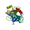
| ||||||||
|---|---|---|---|---|---|---|---|---|---|
| 1 | 
| ||||||||
| Unit cell |
|
-Components
| #1: Protein | Mass: 24668.814 Da / Num. of mol.: 1 / Mutation: Q20Y, E21N, C27V Source method: isolated from a genetically manipulated source Source: (gene. exp.) Homo sapiens (human) / Production host:  |
|---|---|
| #2: Chemical | ChemComp-0G6 /   Type: peptide-like, Peptide-like / Class: Inhibitor / Mass: 453.986 Da / Num. of mol.: 1 / Source method: obtained synthetically / Formula: C21H34ClN6O3 / References: D-Phe-Pro-Arg-CH2Cl Type: peptide-like, Peptide-like / Class: Inhibitor / Mass: 453.986 Da / Num. of mol.: 1 / Source method: obtained synthetically / Formula: C21H34ClN6O3 / References: D-Phe-Pro-Arg-CH2Cl |
| #3: Water | ChemComp-HOH /  Mass: 18.015 Da / Num. of mol.: 82 / Source method: isolated from a natural source / Formula: H2O Mass: 18.015 Da / Num. of mol.: 82 / Source method: isolated from a natural source / Formula: H2O |
| Has protein modification | Y |
| Nonpolymer details | THE UNBOUND FORM OF THE INHIBITOR IS D-PHE-PRO-ARG-CHLOROMETHYLKETONE. UPON REACTION WITH PROTEIN ...THE UNBOUND FORM OF THE INHIBITOR IS D-PHE-PRO-ARG-CHLOROMETH |
| Sequence details | RESIDUES ARE NUMBERED ACCORDING TO THE CHYMOTRYPSIN NUMBERING SYSTEM. THE MOLECULE IS A CHIMERIC ...RESIDUES ARE NUMBERED ACCORDING TO THE CHYMOTRYPS |
-Experimental details
-Experiment
| Experiment | Method: X-RAY DIFFRACTION / Number of used crystals: 1 |
|---|
- Sample preparation
| Crystal | Density Matthews: 2.28 Å3/Da / Density % sol: 46.13 % | ||||||||||||||||||||
|---|---|---|---|---|---|---|---|---|---|---|---|---|---|---|---|---|---|---|---|---|---|
| Crystal grow | pH: 7.8 Details: PROTEIN WAS CRYSTALLIZED FROM 15% PEG 6K, 100 MM HEPES, PH 7.8. | ||||||||||||||||||||
| Crystal grow | *PLUS Temperature: 4 ℃ / Method: vapor diffusion, sitting drop | ||||||||||||||||||||
| Components of the solutions | *PLUS
|
-Data collection
| Diffraction | Mean temperature: 280 K |
|---|---|
| Diffraction source | Source: ROTATING ANODE / Type: RIGAKU RUH2R / Wavelength: 1.5418 |
| Detector | Type: MARRESEARCH / Detector: IMAGE PLATE / Date: May 1, 1995 |
| Radiation | Monochromator: GRAPHITE(002) / Monochromatic (M) / Laue (L): M / Scattering type: x-ray |
| Radiation wavelength | Wavelength: 1.5418 Å / Relative weight: 1 |
| Reflection | Highest resolution: 2.15 Å |
| Reflection | *PLUS Lowest resolution: 8 Å / Num. obs: 10714 / % possible obs: 89.3 % / Rmerge(I) obs: 0.072 / Num. measured all: 28062 |
| Reflection shell | *PLUS Highest resolution: 2.15 Å / Lowest resolution: 2.25 Å / % possible obs: 91.6 % |
- Processing
| Software |
| ||||||||||||||||||||||||||||||||||||||||||||||||||||||||||||
|---|---|---|---|---|---|---|---|---|---|---|---|---|---|---|---|---|---|---|---|---|---|---|---|---|---|---|---|---|---|---|---|---|---|---|---|---|---|---|---|---|---|---|---|---|---|---|---|---|---|---|---|---|---|---|---|---|---|---|---|---|---|
| Refinement | Resolution: 2.15→8 Å / Data cutoff high absF: 1000000 / Data cutoff low absF: 0.001 / Cross valid method: FREE R-VALUE / σ(F): 2
| ||||||||||||||||||||||||||||||||||||||||||||||||||||||||||||
| Refinement step | Cycle: LAST / Resolution: 2.15→8 Å
| ||||||||||||||||||||||||||||||||||||||||||||||||||||||||||||
| Refine LS restraints |
| ||||||||||||||||||||||||||||||||||||||||||||||||||||||||||||
| Xplor file |
| ||||||||||||||||||||||||||||||||||||||||||||||||||||||||||||
| Software | *PLUS Name: X-PLOR / Version: 3.8 / Classification: refinement | ||||||||||||||||||||||||||||||||||||||||||||||||||||||||||||
| Refinement | *PLUS Rfactor obs: 0.185 / Rfactor Rfree: 0.241 | ||||||||||||||||||||||||||||||||||||||||||||||||||||||||||||
| Solvent computation | *PLUS | ||||||||||||||||||||||||||||||||||||||||||||||||||||||||||||
| Displacement parameters | *PLUS | ||||||||||||||||||||||||||||||||||||||||||||||||||||||||||||
| Refine LS restraints | *PLUS Type: x_angle_deg / Dev ideal: 1.2 |
