 Movie
Movie Controller

[English] 日本語
 Yorodumi
- PDB-2ixd: Crystal structure of the putative deacetylase BC1534 from Bacillu... -
+ Open data

- Basic information
| Entry | Database: PDB / ID: 2ixd | ||||||
|---|---|---|---|---|---|---|---|
| Title | Crystal structure of the putative deacetylase BC1534 from Bacillus cereus | ||||||
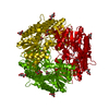
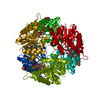
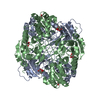
 Components Components | LMBE-RELATED PROTEIN | ||||||
 Keywords Keywords | HYDROLASE / HEXAMER / DEACETYLASE / ROSSMANN FOLD / ZINC-DEPENDENT METALLOENZYME | ||||||
| Function / homology |  Function and homology information Function and homology informationbacillithiol biosynthetic process / hydrolase activity, acting on carbon-nitrogen (but not peptide) bonds, in linear amides / deacetylase activity / Hydrolases; Acting on carbon-nitrogen bonds, other than peptide bonds; In linear amides / metal ion binding Similarity search - Function | ||||||
| Biological species |  | ||||||
| Method |  X-RAY DIFFRACTION / SYNCHROTRON / MOLECULAR REPLACEMENT / Resolution: 1.8 Å X-RAY DIFFRACTION / SYNCHROTRON / MOLECULAR REPLACEMENT / Resolution: 1.8 Å | ||||||
 Authors Authors | Fadouloglou, V.E. / Bouriotis, V. / Kokkinidis, M. | ||||||
 Citation Citation | Journal: FEBS J. / Year: 2007 Title: Crystal Structure of the Bczbp, a Zinc-Binding Protein from Bacillus Cereus Authors: Fadouloglou, V.E. / Deli, A. / Glykos, N.M. / Psylinakis, E. / Bouriotis, V. / Kokkinidis, M. #1: Journal: Acta Crystallogr.,Sect.F / Year: 2006 Title: Purification, Crystallization and Preliminary Characterization of a Putative Lmbe-Like Deacetylase from Bacillus Cereus Authors: Fadouloglou, V.E. / Kotsifaki, D. / Gazi, A.D. / Fellas, G. / Meramveliotaki, C. / Deli, A. / Psylinakis, E. / Bouriotis, V. / Kokkinidis, M. | ||||||
| History |
|
- Structure visualization
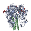
| Structure viewer | Molecule: MolmilJmol/JSmol |
|---|
- Downloads & links
-Download
| PDBx/mmCIF format | 2ixd.cif.gz | 205.5 KB | Display | PDBx/mmCIF format |
|---|---|---|---|---|
| PDB format | pdb2ixd.ent.gz | 167.1 KB | Display | PDB format |
| PDBx/mmJSON format | 2ixd.json.gz | Tree view | PDBx/mmJSON format | |
| Others |  Other downloads Other downloads |
-Validation report
| Arichive directory | https://data.pdbj.org/pub/pdb/validation_reports/ix/2ixdftp://data.pdbj.org/pub/pdb/validation_reports/ix/2ixd | HTTPS FTP |
|---|
-Related structure data
| Related structure data | 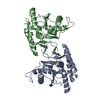 1uanS S: Starting model for refinement |
|---|---|
| Similar structure data |

-Links
 PDBj
PDBj- Assembly
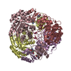
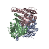
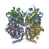
| Deposited unit | 
| ||||||||
|---|---|---|---|---|---|---|---|---|---|
| 1 | 
| ||||||||
| Unit cell |
| ||||||||
| Components on special symmetry positions |
| ||||||||
| Noncrystallographic symmetry (NCS) | NCS oper: (Code: given Matrix: (0.94418, -0.32914, 0.01347), Vector: |
-Components
| #1: Protein | Mass: 27256.383 Da / Num. of mol.: 2 Source method: isolated from a genetically manipulated source Source: (gene. exp.) #2: Chemical |   Mass: 65.409 Da / Num. of mol.: 2 / Source method: obtained synthetically / Formula: Zn Mass: 65.409 Da / Num. of mol.: 2 / Source method: obtained synthetically / Formula: Zn#3: Chemical |   Mass: 59.044 Da / Num. of mol.: 2 / Source method: obtained synthetically / Formula: C2H3O2 Mass: 59.044 Da / Num. of mol.: 2 / Source method: obtained synthetically / Formula: C2H3O2#4: Water | ChemComp-HOH / |  Mass: 18.015 Da / Num. of mol.: 471 / Source method: isolated from a natural source / Formula: H2O Mass: 18.015 Da / Num. of mol.: 471 / Source method: isolated from a natural source / Formula: H2O |
|---|
-Experimental details
-Experiment
| Experiment | Method: X-RAY DIFFRACTION / Number of used crystals: 1 |
|---|
- Sample preparation
| Crystal | Density Matthews: 2.2 Å3/Da / Density % sol: 44 % |
|---|---|
| Crystal grow | pH: 5 / Details: 14-26% MPD, 100 MM ACETATE BUFFER, PH 5.0 |
-Data collection
| Diffraction | Mean temperature: 100 K |
|---|---|
| Diffraction source | Source: SYNCHROTRON / Site: EMBL/DESY, HAMBURG  / Beamline: X12 / Wavelength: 1.2821 / Beamline: X12 / Wavelength: 1.2821 |
| Detector | Type: MARRESEARCH / Detector: CCD / Date: Nov 2, 2005 |
| Radiation | Protocol: SINGLE WAVELENGTH / Monochromatic (M) / Laue (L): M / Scattering type: x-ray |
| Radiation wavelength | Wavelength: 1.2821 Å / Relative weight: 1 |
| Reflection | Resolution: 1.8→55.8 Å / Num. obs: 40471 / % possible obs: 92.3 % / Observed criterion σ(I): 0 / Redundancy: 7.3 % / Biso Wilson estimate: 23.4 Å2 / Rmerge(I) obs: 0.06 / Net I/σ(I): 18.3 |
| Reflection shell | Resolution: 1.8→1.9 Å / Redundancy: 6.3 % / Rmerge(I) obs: 0.5 / Mean I/σ(I) obs: 3.5 / % possible all: 65.4 |
- Processing
| Software |
| ||||||||||||||||||||||||||||||||||||||||||||||||||||||||||||||||||||||||||||||||||||||||||||||||||||||||||||||||||||||||||||||||||||||||||||||||||||||||||||||||||||||||||||||||||||||
|---|---|---|---|---|---|---|---|---|---|---|---|---|---|---|---|---|---|---|---|---|---|---|---|---|---|---|---|---|---|---|---|---|---|---|---|---|---|---|---|---|---|---|---|---|---|---|---|---|---|---|---|---|---|---|---|---|---|---|---|---|---|---|---|---|---|---|---|---|---|---|---|---|---|---|---|---|---|---|---|---|---|---|---|---|---|---|---|---|---|---|---|---|---|---|---|---|---|---|---|---|---|---|---|---|---|---|---|---|---|---|---|---|---|---|---|---|---|---|---|---|---|---|---|---|---|---|---|---|---|---|---|---|---|---|---|---|---|---|---|---|---|---|---|---|---|---|---|---|---|---|---|---|---|---|---|---|---|---|---|---|---|---|---|---|---|---|---|---|---|---|---|---|---|---|---|---|---|---|---|---|---|---|---|
| Refinement | Method to determine structure: MOLECULAR REPLACEMENT Starting model: PDB ENTRY 1UAN Resolution: 1.8→55.81 Å / Cor.coef. Fo:Fc: 0.955 / Cor.coef. Fo:Fc free: 0.945 / SU B: 2.747 / SU ML: 0.086 / Cross valid method: THROUGHOUT / ESU R: 0.149 / ESU R Free: 0.13 / Stereochemistry target values: MAXIMUM LIKELIHOOD / Details: HYDROGENS HAVE BEEN ADDED IN THE RIDING POSITIONS.
| ||||||||||||||||||||||||||||||||||||||||||||||||||||||||||||||||||||||||||||||||||||||||||||||||||||||||||||||||||||||||||||||||||||||||||||||||||||||||||||||||||||||||||||||||||||||
| Solvent computation | Ion probe radii: 0.8 Å / Shrinkage radii: 0.8 Å / VDW probe radii: 1.4 Å / Solvent model: BABINET MODEL WITH MASK | ||||||||||||||||||||||||||||||||||||||||||||||||||||||||||||||||||||||||||||||||||||||||||||||||||||||||||||||||||||||||||||||||||||||||||||||||||||||||||||||||||||||||||||||||||||||
| Displacement parameters | Biso mean: 15.2 Å2
| ||||||||||||||||||||||||||||||||||||||||||||||||||||||||||||||||||||||||||||||||||||||||||||||||||||||||||||||||||||||||||||||||||||||||||||||||||||||||||||||||||||||||||||||||||||||
| Refinement step | Cycle: LAST / Resolution: 1.8→55.81 Å
| ||||||||||||||||||||||||||||||||||||||||||||||||||||||||||||||||||||||||||||||||||||||||||||||||||||||||||||||||||||||||||||||||||||||||||||||||||||||||||||||||||||||||||||||||||||||
| Refine LS restraints |
|