- PDB-4km9: Crystal structure of human Suppressor of Fused -
+
Open data
ID or keywords:
Loading...
-
Basic information
Entry
Database: PDB / ID: 4km9
Title
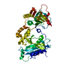
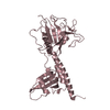
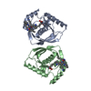
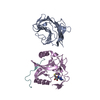
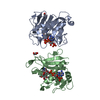
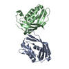
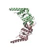
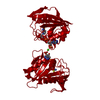
Crystal structure of human Suppressor of Fused
Components
Suppressor of fused homolog
Keywords
PROTEIN BINDING / Suppressor of Fused
Function / homology
Function and homology information
smoothened signaling pathway involved in ventral spinal cord interneuron specification / smoothened signaling pathway involved in spinal cord motor neuron cell fate specification / positive regulation of cellular response to drug / maintenance of protein localization in organelle / GLI-SUFU complex / coronary vasculature development / ventricular septum development / aorta development / dorsal/ventral neural tube patterning / negative regulation of protein import into nucleus ...smoothened signaling pathway involved in ventral spinal cord interneuron specification / smoothened signaling pathway involved in spinal cord motor neuron cell fate specification / positive regulation of cellular response to drug / maintenance of protein localization in organelle / GLI-SUFU complex / coronary vasculature development / ventricular septum development / aorta development / dorsal/ventral neural tube patterning / negative regulation of protein import into nucleus / skin development / heart looping / ciliary base / spermatid development / negative regulation of osteoblast differentiation / negative regulation of ubiquitin-dependent protein catabolic process / Hedgehog 'off' state / ciliary tip / neural tube closure / negative regulation of smoothened signaling pathway / protein sequestering activity / Degradation of GLI1 by the proteasome / Hedgehog 'on' state / beta-catenin binding / Degradation of GLI2 by the proteasome / GLI3 is processed to GLI3R by the proteasome / transcription corepressor activity / cilium / regulation of DNA-templated transcription / protein kinase binding / negative regulation of transcription by RNA polymerase II / signal transduction / nucleoplasm / nucleus / cytosol / cytoplasm Similarity search - Function
Sufu, C-terminal domain / Suppressor of fused / Suppressor of fused, eukaryotic / Suppressor of fused C-terminal / Suppressor of fused, N-terminal / Suppressor of fused, C-terminal domain superfamily / Suppressor of Fused Gli/Ci N terminal binding domain / Suppressor of fused-like domain / Suppressor of fused protein (SUFU) / Gyrase A; domain 2 ...Sufu, C-terminal domain / Suppressor of fused / Suppressor of fused, eukaryotic / Suppressor of fused C-terminal / Suppressor of fused, N-terminal / Suppressor of fused, C-terminal domain superfamily / Suppressor of Fused Gli/Ci N terminal binding domain / Suppressor of fused-like domain / Suppressor of fused protein (SUFU) / Gyrase A; domain 2 / 2-Layer Sandwich / Alpha Beta Similarity search - Domain/homology
Mass: 54451.066 Da / Num. of mol.: 1 Source method: isolated from a genetically manipulated source Source: (gene. exp.) Homo sapiens (human) / Gene: SUFU, UNQ650/PRO1280 / Cell (production host): High Five / Production host: Trichoplusia ni (cabbage looper) / References: UniProt: Q9UMX1
Resolution: 3.19→50 Å / Cor.coef. Fo:Fc: 0.918 / Cor.coef. Fo:Fc free: 0.849 / Occupancy max: 1 / Occupancy min: 0.2 / SU B: 59.121 / SU ML: 0.471 / Cross valid method: THROUGHOUT / σ(F): 0 / ESU R Free: 0.589 / Stereochemistry target values: MAXIMUM LIKELIHOOD Details: HYDROGENS HAVE BEEN ADDED IN THE RIDING POSITIONS U VALUES: RESIDUAL ONLY
Rfactor
Num. reflection
% reflection
Selection details
Rfree
0.3053
426
4.8 %
RANDOM
Rwork
0.2434
-
-
-
obs
0.2462
8939
98.76 %
-
Solvent computation
Ion probe radii: 0.8 Å / Shrinkage radii: 0.8 Å / VDW probe radii: 1.4 Å / Solvent model: MASK
In the structure databanks used in Yorodumi, some data are registered as the other names, "COVID-19 virus" and "2019-nCoV". Here are the details of the virus and the list of structure data.
Jan 31, 2019. EMDB accession codes are about to change! (news from PDBe EMDB page)
EMDB accession codes are about to change! (news from PDBe EMDB page)
The allocation of 4 digits for EMDB accession codes will soon come to an end. Whilst these codes will remain in use, new EMDB accession codes will include an additional digit and will expand incrementally as the available range of codes is exhausted. The current 4-digit format prefixed with “EMD-” (i.e. EMD-XXXX) will advance to a 5-digit format (i.e. EMD-XXXXX), and so on. It is currently estimated that the 4-digit codes will be depleted around Spring 2019, at which point the 5-digit format will come into force.
The EM Navigator/Yorodumi systems omit the EMD- prefix.
Related info.:Q: What is EMD? / ID/Accession-code notation in Yorodumi/EM Navigator
Yorodumi is a browser for structure data from EMDB, PDB, SASBDB, etc.
This page is also the successor to EM Navigator detail page, and also detail information page/front-end page for Omokage search.
The word "yorodu" (or yorozu) is an old Japanese word meaning "ten thousand". "mi" (miru) is to see.
Related info.:EMDB / PDB / SASBDB / Comparison of 3 databanks / Yorodumi Search / Aug 31, 2016. New EM Navigator & Yorodumi / Yorodumi Papers / Jmol/JSmol / Function and homology information / Changes in new EM Navigator and Yorodumi
 Movie
Movie Controller


 Open data
 Basic information
 Components
 Keywords
 Function and homology information
 Homo sapiens (human)
 X-RAY DIFFRACTION /
  Authors
 Citation
 Structure visualization
 Downloads & links
 Other downloads
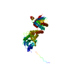


 PDBj
PDBj

 Assembly
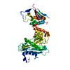
 Trichoplusia ni (cabbage looper) / References: UniProt: Q9UMX1
 Mass: 18.015 Da / Num. of mol.: 7 / Source method: isolated from a natural source / Formula: H2O
 Sample preparation
 / Beamline: BL17U / Wavelength: 1 Å
 Processing