 Movie
Movie Controller

[English] 日本語
 Yorodumi
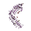
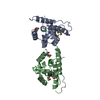
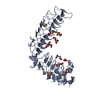
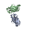
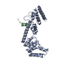
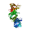
- PDB-1gml: crystal structure of the mouse CCT gamma apical domain (triclinic) -
+ Open data

- Basic information
| Entry | Database: PDB / ID: 1gml | ||||||
|---|---|---|---|---|---|---|---|
| Title | crystal structure of the mouse CCT gamma apical domain (triclinic) | ||||||
 Components Components | T-COMPLEX PROTEIN 1 SUBUNIT GAMMA | ||||||
 Keywords Keywords | CHAPERONE / CHAPERONIN / ACTIN / TUBULIN | ||||||
| Function / homology |  Function and homology information Function and homology informationAssociation of TriC/CCT with target proteins during biosynthesis / BBSome-mediated cargo-targeting to cilium / Cooperation of PDCL (PhLP1) and TRiC/CCT in G-protein beta folding / zona pellucida receptor complex / chaperonin-containing T-complex / binding of sperm to zona pellucida / Hydrolases; Acting on acid anhydrides; In phosphorus-containing anhydrides / positive regulation of telomere maintenance via telomerase / protein folding chaperone / ATP-dependent protein folding chaperone ...Association of TriC/CCT with target proteins during biosynthesis / BBSome-mediated cargo-targeting to cilium / Cooperation of PDCL (PhLP1) and TRiC/CCT in G-protein beta folding / zona pellucida receptor complex / chaperonin-containing T-complex / binding of sperm to zona pellucida / Hydrolases; Acting on acid anhydrides; In phosphorus-containing anhydrides / positive regulation of telomere maintenance via telomerase / protein folding chaperone / ATP-dependent protein folding chaperone / : / myelin sheath / protein folding / cell body / microtubule / protein stabilization / ATP hydrolysis activity / ATP binding / metal ion binding Similarity search - Function | ||||||
| Biological species |  | ||||||
| Method |  X-RAY DIFFRACTION / SYNCHROTRON / MOLECULAR REPLACEMENT / Resolution: 2.2 Å X-RAY DIFFRACTION / SYNCHROTRON / MOLECULAR REPLACEMENT / Resolution: 2.2 Å | ||||||
 Authors Authors | Pappenberger, G. / Wilsher, J.A. / Roe, S.M. / Willison, K.R. / Pearl, L.H. | ||||||
 Citation Citation | Journal: J.Mol.Biol. / Year: 2002 Title: Crystal Structure of the Cct Gamma Apical Domain:: Implications for Substrate Binding to the Eukaryotic Cytosolic Chaperonin Authors: Pappenberger, G. / Wilsher, J.A. / Roe, S.M. / Counsell, D.J. / Willison, K.R. / Pearl, L.H. | ||||||
| History |
|
- Structure visualization
| Structure viewer | Molecule: MolmilJmol/JSmol |
|---|
- Downloads & links
-Download
| PDBx/mmCIF format | 1gml.cif.gz | 145.1 KB | Display | PDBx/mmCIF format |
|---|---|---|---|---|
| PDB format | pdb1gml.ent.gz | 114.4 KB | Display | PDB format |
| PDBx/mmJSON format | 1gml.json.gz | Tree view | PDBx/mmJSON format | |
| Others |  Other downloads Other downloads |
-Validation report
| Arichive directory | https://data.pdbj.org/pub/pdb/validation_reports/gm/1gmlftp://data.pdbj.org/pub/pdb/validation_reports/gm/1gml | HTTPS FTP |
|---|
-Related structure data
| Related structure data |  1gn1C  1e0rS S: Starting model for refinement C: citing same article ( |
|---|---|
| Similar structure data |


-Links
 PDBj
PDBj


- Assembly
| Deposited unit | 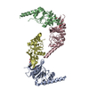
| ||||||||
|---|---|---|---|---|---|---|---|---|---|
| 1 | 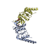
| ||||||||
| 2 | 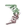
| ||||||||
| Unit cell |
| ||||||||
| Details | THE KNOWN BIOLOGICALLY SIGNIFICANT OLIGOMERIZATION STATEOF THE MOLECULE IS THE MONOMER. TOGETHER WITH THE SEVEN OTHER SUBUNITS OF CCT, IT IS PART OF A DOUBLE TOROIDAL QUATERNARY STRUCTURE OF 2X8 SUBUNITS. |
-Components
| #1: Protein | Mass: 20644.701 Da / Num. of mol.: 4 / Fragment: APICAL DOMAIN, RESIDUES 210-380 Source method: isolated from a genetically manipulated source Source: (gene. exp.)  #2: Chemical | ChemComp-GOL /   Mass: 92.094 Da / Num. of mol.: 4 / Source method: obtained synthetically / Formula: C3H8O3 Mass: 92.094 Da / Num. of mol.: 4 / Source method: obtained synthetically / Formula: C3H8O3#3: Water | ChemComp-HOH / |  Mass: 18.015 Da / Num. of mol.: 645 / Source method: isolated from a natural source / Formula: H2O Mass: 18.015 Da / Num. of mol.: 645 / Source method: isolated from a natural source / Formula: H2OHas protein modification | Y | |
|---|
-Experimental details
-Experiment
| Experiment | Method: X-RAY DIFFRACTION / Number of used crystals: 1 |
|---|
- Sample preparation
| Crystal | Density Matthews: 2.6 Å3/Da / Density % sol: 52 % | ||||||||||||||||||||||||||||||||||||||||||||||||||||||||||||||||||||||
|---|---|---|---|---|---|---|---|---|---|---|---|---|---|---|---|---|---|---|---|---|---|---|---|---|---|---|---|---|---|---|---|---|---|---|---|---|---|---|---|---|---|---|---|---|---|---|---|---|---|---|---|---|---|---|---|---|---|---|---|---|---|---|---|---|---|---|---|---|---|---|---|
| Crystal grow | Method: vapor diffusion, hanging drop / pH: 8 Details: CRYSTALS GROWN BY HANGING DROP METHOD USING A 1:1 MIXTURE OF 12MG/ML PROTEIN, 400MM NACL, 20% GLYCEROL, 8MM TRIS PH8.0, 0.4MM EDTA AND 100MM TRIS PH8.0, 200MM NACL, 10MM MG(OAC)2, pH 8.00 | ||||||||||||||||||||||||||||||||||||||||||||||||||||||||||||||||||||||
| Crystal grow | *PLUS Temperature: 14 ℃ / Method: vapor diffusion, hanging drop | ||||||||||||||||||||||||||||||||||||||||||||||||||||||||||||||||||||||
| Components of the solutions | *PLUS
|
-Data collection
| Diffraction | Mean temperature: 100 K |
|---|---|
| Diffraction source | Source: SYNCHROTRON / Site: SRS  / Beamline: PX9.6 / Wavelength: 0.87 / Beamline: PX9.6 / Wavelength: 0.87 |
| Detector | Type: ADSC CCD / Detector: CCD / Date: Mar 3, 2001 |
| Radiation | Protocol: SINGLE WAVELENGTH / Monochromatic (M) / Laue (L): M / Scattering type: x-ray |
| Radiation wavelength | Wavelength: 0.87 Å / Relative weight: 1 |
| Reflection | Resolution: 2.2→30 Å / Num. obs: 39106 / % possible obs: 93 % / Redundancy: 1.9 % / Biso Wilson estimate: 31.5 Å2 / Rmerge(I) obs: 0.058 / Net I/σ(I): 8 |
| Reflection shell | Resolution: 2.2→2.31 Å / Redundancy: 1.8 % / Rmerge(I) obs: 0.15 / Mean I/σ(I) obs: 4.2 / % possible all: 92.6 |
| Reflection | *PLUS Lowest resolution: 29 Å |
| Reflection shell | *PLUS % possible obs: 92.6 % / Rmerge(I) obs: 0.15 |
- Processing
| Software |
| ||||||||||||||||||||||||||||||||||||||||||||||||||||||||||||||||||||||||||||||||
|---|---|---|---|---|---|---|---|---|---|---|---|---|---|---|---|---|---|---|---|---|---|---|---|---|---|---|---|---|---|---|---|---|---|---|---|---|---|---|---|---|---|---|---|---|---|---|---|---|---|---|---|---|---|---|---|---|---|---|---|---|---|---|---|---|---|---|---|---|---|---|---|---|---|---|---|---|---|---|---|---|---|
| Refinement | Method to determine structure: MOLECULAR REPLACEMENT Starting model: PDB ENTRY 1E0R Resolution: 2.2→28.95 Å / Rfactor Rfree error: 0.005 / Data cutoff high absF: 1072988.9 / Isotropic thermal model: RESTRAINED / Cross valid method: THROUGHOUT / σ(F): 0 Stereochemistry target values: MAXIMUM LIKELIHOOD USING AMPLITUDES Details: NO DENSITY WAS FOUND FOR THE LOOPS A249 - A262 B249 - B261 C249 - C262 D249 - D268 NO DENSITY WAS FOUND FOR SEVERAL SIDECHAINS. THESE WERE TRUNCATED AT CB.
| ||||||||||||||||||||||||||||||||||||||||||||||||||||||||||||||||||||||||||||||||
| Solvent computation | Solvent model: FLAT MODEL / Bsol: 59.9464 Å2 / ksol: 0.322875 e/Å3 | ||||||||||||||||||||||||||||||||||||||||||||||||||||||||||||||||||||||||||||||||
| Displacement parameters | Biso mean: 41.9 Å2
| ||||||||||||||||||||||||||||||||||||||||||||||||||||||||||||||||||||||||||||||||
| Refine analyze |
| ||||||||||||||||||||||||||||||||||||||||||||||||||||||||||||||||||||||||||||||||
| Refinement step | Cycle: LAST / Resolution: 2.2→28.95 Å
| ||||||||||||||||||||||||||||||||||||||||||||||||||||||||||||||||||||||||||||||||
| Refine LS restraints |
| ||||||||||||||||||||||||||||||||||||||||||||||||||||||||||||||||||||||||||||||||
| Refine LS restraints NCS | Rms dev Biso : 0.2 Å2 / Weight position: 2000 | ||||||||||||||||||||||||||||||||||||||||||||||||||||||||||||||||||||||||||||||||
| LS refinement shell | Resolution: 2.2→2.34 Å / Rfactor Rfree error: 0.022 / Total num. of bins used: 6
| ||||||||||||||||||||||||||||||||||||||||||||||||||||||||||||||||||||||||||||||||
| Xplor file |
| ||||||||||||||||||||||||||||||||||||||||||||||||||||||||||||||||||||||||||||||||
| Refine LS restraints | *PLUS
| ||||||||||||||||||||||||||||||||||||||||||||||||||||||||||||||||||||||||||||||||
| LS refinement shell | *PLUS Rfactor obs: 0.373 |
