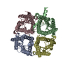
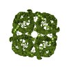
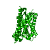
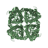
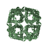
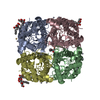
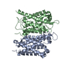
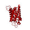
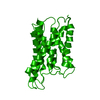
ジャーナル: Nature / 年: 2000 タイトル: Structural determinants of water permeation through aquaporin-1. 著者: K Murata / K Mitsuoka / T Hirai / T Walz / P Agre / J B Heymann / A Engel / Y Fujiyoshi / 要旨: Human red cell AQP1 is the first functionally defined member of the aquaporin family of membrane water channels. Here we describe an atomic model of AQP1 at 3.8A resolution from electron ...Human red cell AQP1 is the first functionally defined member of the aquaporin family of membrane water channels. Here we describe an atomic model of AQP1 at 3.8A resolution from electron crystallographic data. Multiple highly conserved amino-acid residues stabilize the novel fold of AQP1. The aqueous pathway is lined with conserved hydrophobic residues that permit rapid water transport, whereas the water selectivity is due to a constriction of the pore diameter to about 3 A over a span of one residue. The atomic model provides a possible molecular explanation to a longstanding puzzle in physiology-how membranes can be freely permeable to water but impermeable to protons.
履歴
登録
2000年9月7日
登録サイト: RCSB / 処理サイト: RCSB
改定 1.0
2000年10月18日
Provider: repository / タイプ: Initial release
改定 1.1
2008年4月27日
Group: Version format compliance
改定 1.2
2011年7月13日
Group: Derived calculations / Version format compliance
解像度: 3.8 Å / 解像度の算出法: DIFFRACTION PATTERN/LAYERLINES
精密化
解像度: 3.8→6 Å / σ(F): 1 / σ(I): 0 / 立体化学のターゲット値: Engh & Huber 詳細: For refinement with phase restraint, we used the structure factors merged from both electron diffraction patterns and images. Thus in this file we included the structure factors with phases ...詳細: For refinement with phase restraint, we used the structure factors merged from both electron diffraction patterns and images. Thus in this file we included the structure factors with phases used in the refinement. The observed phases were labelled as calculated phases here.
 ムービー
ムービー コントローラー


 データを開く
 基本情報
基本情報 要素
要素 キーワード
キーワード 機能・相同性情報
 Homo sapiens (ヒト)
 データ登録者
 引用
引用
 構造の表示
構造の表示 ダウンロードとリンク
 その他のダウンロード
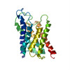


 PDBj
PDBj

 集合体
集合体
 試料調製
試料調製 FIELD EMISSION GUN / 加速電圧: 300 kV / 照射モード: FLOOD BEAM
 解析
解析