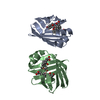
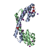
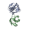
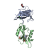
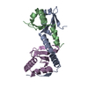
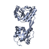
Mass: 12250.814 Da / Num. of mol.: 2 Source method: isolated from a genetically manipulated source Source: (gene. exp.) Burkholderia xenovorans (bacteria) / Strain: LB400 / Plasmid: PEBRE12 / Production host: Escherichia coli (E. coli) References: UniProt: P37332, Oxidoreductases; Acting on paired donors, with incorporation or reduction of molecular oxygen; With NADH or NADPH as one donor, and incorporation of two atoms of oxygen into the other donor
Resolution: 1.6→26 Å / Num. obs: 34971 / % possible obs: 98.9 % / Observed criterion σ(I): 0 / Redundancy: 4.95 % / Biso Wilson estimate: 14.5 Å2 / Rmerge(I) obs: 0.068 / Net I/σ(I): 9.6
Reflection shell
Resolution: 1.6→1.66 Å / Redundancy: 4.91 % / Rmerge(I) obs: 0.412 / Mean I/σ(I) obs: 2.2 / % possible all: 97.6
Reflection shell
*PLUS
% possible obs: 97.6 %
-
Processing
Software
Name
Classification
MLPHARE
phasing
CNS
refinement
DENZO
datareduction
SCALEPACK
datascaling
Refinement
Method to determine structure: FE MAD / Resolution: 1.6→26 Å / Rfactor Rfree error: 0.005 / Isotropic thermal model: RESTRAINED / Cross valid method: THROUGHOUT / σ(F): 0 / Stereochemistry target values: ENGH & HUBER Details: THE GEOMETRY WITHIN THE FE-S CLUSTER AND THE BONDS WITH THE PROTEIN LIGANDS WERE RESTRAINED WITH FORCE CONSTANTS OF: 200 KCAL MOLE^-1 ANG.^2 APPLIED TO FE-S (TARGET=2.28 ANG.), FE-ND1 (2.05 ...Details: THE GEOMETRY WITHIN THE FE-S CLUSTER AND THE BONDS WITH THE PROTEIN LIGANDS WERE RESTRAINED WITH FORCE CONSTANTS OF: 200 KCAL MOLE^-1 ANG.^2 APPLIED TO FE-S (TARGET=2.28 ANG.), FE-ND1 (2.05 ANG.), AND FE-FE (2.68 ANG.) DISTANCES; 40 KCAL MOLE^-1 RAD^-2 APPLIED TO ANGLES S-FE-ND1 (115 DEG.), S-FE-S (105 DEG.), FE-S-FE (75 DEG.), ND1-FE-ND1 (90 DEG.), FE-ND1-CG (108 DEG.), AND FE-S-CB (109.5 DEG.) REFINEMENT TARGET EQUALS MAXIMUM LIKELIHOOD TARGET USING AMPLITUDES
In the structure databanks used in Yorodumi, some data are registered as the other names, "COVID-19 virus" and "2019-nCoV". Here are the details of the virus and the list of structure data.
Jan 31, 2019. EMDB accession codes are about to change! (news from PDBe EMDB page)
EMDB accession codes are about to change! (news from PDBe EMDB page)
The allocation of 4 digits for EMDB accession codes will soon come to an end. Whilst these codes will remain in use, new EMDB accession codes will include an additional digit and will expand incrementally as the available range of codes is exhausted. The current 4-digit format prefixed with “EMD-” (i.e. EMD-XXXX) will advance to a 5-digit format (i.e. EMD-XXXXX), and so on. It is currently estimated that the 4-digit codes will be depleted around Spring 2019, at which point the 5-digit format will come into force.
The EM Navigator/Yorodumi systems omit the EMD- prefix.
Related info.:Q: What is EMD? / ID/Accession-code notation in Yorodumi/EM Navigator
Yorodumi is a browser for structure data from EMDB, PDB, SASBDB, etc.
This page is also the successor to EM Navigator detail page, and also detail information page/front-end page for Omokage search.
The word "yorodu" (or yorozu) is an old Japanese word meaning "ten thousand". "mi" (miru) is to see.
Related info.:EMDB / PDB / SASBDB / Comparison of 3 databanks / Yorodumi Search / Aug 31, 2016. New EM Navigator & Yorodumi / Yorodumi Papers / Jmol/JSmol / Function and homology information / Changes in new EM Navigator and Yorodumi
 Movie
Movie Controller

 Yorodumi
 Open data
 Basic information
 Components
 Keywords
 Function and homology information
 Burkholderia xenovorans (bacteria)
 X-RAY DIFFRACTION /
  Authors
 Citation
 Structure visualization
 Downloads & links
 Other downloads
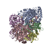


 PDBj
PDBj


 Assembly
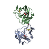

 Mass: 175.820 Da / Num. of mol.: 2 / Source method: obtained synthetically / Formula: Fe2S2
 Mass: 92.094 Da / Num. of mol.: 3 / Source method: obtained synthetically / Formula: C3H8O3
 Mass: 18.015 Da / Num. of mol.: 314 / Source method: isolated from a natural source / Formula: H2O
 Sample preparation
 / Beamline: 14-BM-D / Wavelength: 0.9537,1.742,1.737
 Processing