 Movie
Movie Controller

[English] 日本語
 Yorodumi
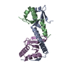
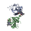
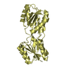
- PDB-3re1: Crystal structure of uroporphyrinogen III synthase from Pseudomon... -
+ Open data

- Basic information
| Entry | Database: PDB / ID: 3re1 | ||||||
|---|---|---|---|---|---|---|---|
| Title | Crystal structure of uroporphyrinogen III synthase from Pseudomonas syringae pv. tomato DC3000 | ||||||
 Components Components | Uroporphyrinogen-III synthetase | ||||||
 Keywords Keywords | LYASE / HemD-like family / uroporphyrinogen III synthase / HMB | ||||||
| Function / homology |  Function and homology information Function and homology informationuroporphyrinogen-III synthase / uroporphyrinogen-III synthase activity / uroporphyrinogen III biosynthetic process / : Similarity search - Function | ||||||
| Biological species |  Pseudomonas syringae pv. tomato (bacteria) Pseudomonas syringae pv. tomato (bacteria) | ||||||
| Method |  X-RAY DIFFRACTION / SYNCHROTRON / SAD / Resolution: 2.5 Å X-RAY DIFFRACTION / SYNCHROTRON / SAD / Resolution: 2.5 Å | ||||||
 Authors Authors | Chang, W.R. / Li, M. / Peng, S.X. | ||||||
 Citation Citation | Journal: Biochem.Biophys.Res.Commun. / Year: 2011 Title: Crystal structure of uroporphyrinogen III synthase from Pseudomonas syringae pv. tomato DC3000 Authors: Peng, S.X. / Zhang, H. / Gao, Y. / Pan, X. / Cao, P. / Li, M. / Chang, W.R. | ||||||
| History |
|
- Structure visualization
| Structure viewer | Molecule: MolmilJmol/JSmol |
|---|
- Downloads & links
-Download
| PDBx/mmCIF format | 3re1.cif.gz | 113.3 KB | Display | PDBx/mmCIF format |
|---|---|---|---|---|
| PDB format | pdb3re1.ent.gz | 87.5 KB | Display | PDB format |
| PDBx/mmJSON format | 3re1.json.gz | Tree view | PDBx/mmJSON format | |
| Others |  Other downloads Other downloads |
-Validation report
| Arichive directory | https://data.pdbj.org/pub/pdb/validation_reports/re/3re1ftp://data.pdbj.org/pub/pdb/validation_reports/re/3re1 | HTTPS FTP |
|---|
-Related structure data
| Similar structure data |
|---|


-Links
 PDBj
PDBj- Assembly
| Deposited unit | 
| ||||||||
|---|---|---|---|---|---|---|---|---|---|
| 1 | 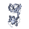
| ||||||||
| 2 | 
| ||||||||
| Unit cell |
|
-Components
| #1: Protein | Mass: 29322.516 Da / Num. of mol.: 2 Source method: isolated from a genetically manipulated source Source: (gene. exp.) Pseudomonas syringae pv. tomato (bacteria)Strain: DC3000 / Gene: hemD / Plasmid: pHAT2 / Production host: #2: Water | ChemComp-HOH / |  Mass: 18.015 Da / Num. of mol.: 275 / Source method: isolated from a natural source / Formula: H2O Mass: 18.015 Da / Num. of mol.: 275 / Source method: isolated from a natural source / Formula: H2O |
|---|
-Experimental details
-Experiment
| Experiment | Method: X-RAY DIFFRACTION / Number of used crystals: 1 |
|---|
- Sample preparation
| Crystal | Density Matthews: 2.42 Å3/Da / Density % sol: 49.17 % |
|---|---|
| Crystal grow | Temperature: 281 K / Method: vapor diffusion, sitting drop / pH: 7.2 Details: 20% PEG3350, 0.2M tri-Na citrate, pH 7.2, VAPOR DIFFUSION, SITTING DROP, temperature 281K |
-Data collection
| Diffraction | Mean temperature: 100 K |
|---|---|
| Diffraction source | Source: SYNCHROTRON / Site: SSRF  / Beamline: BL17U / Wavelength: 0.97916 Å / Beamline: BL17U / Wavelength: 0.97916 Å |
| Detector | Type: MARMOSAIC 225 mm CCD / Detector: CCD / Date: Oct 13, 2010 |
| Radiation | Protocol: SINGLE WAVELENGTH / Monochromatic (M) / Laue (L): M / Scattering type: x-ray |
| Radiation wavelength | Wavelength: 0.97916 Å / Relative weight: 1 |
| Reflection | Resolution: 2.5→50 Å / Num. all: 19467 / Num. obs: 19409 / % possible obs: 99.7 % / Observed criterion σ(F): 2 / Observed criterion σ(I): 2 / Biso Wilson estimate: 34.16 Å2 |
| Reflection shell | Resolution: 2.5→2.59 Å / % possible all: 99.2 |
- Processing
| Software |
| ||||||||||||||||||||||||||||||||||||||||||||||||||||||||
|---|---|---|---|---|---|---|---|---|---|---|---|---|---|---|---|---|---|---|---|---|---|---|---|---|---|---|---|---|---|---|---|---|---|---|---|---|---|---|---|---|---|---|---|---|---|---|---|---|---|---|---|---|---|---|---|---|---|
| Refinement | Method to determine structure: SAD / Resolution: 2.5→30 Å / Occupancy max: 1 / Occupancy min: 1 / FOM work R set: 0.7836 / SU ML: 0.34 / σ(F): 0 / Phase error: 27.88 / Stereochemistry target values: ML / Details: The structure was refined also with CNS 1.2.
| ||||||||||||||||||||||||||||||||||||||||||||||||||||||||
| Solvent computation | Shrinkage radii: 0.95 Å / VDW probe radii: 1.2 Å / Solvent model: FLAT BULK SOLVENT MODEL / Bsol: 31.765 Å2 / ksol: 0.301 e/Å3 | ||||||||||||||||||||||||||||||||||||||||||||||||||||||||
| Displacement parameters | Biso max: 96 Å2 / Biso mean: 45.608 Å2 / Biso min: 5.54 Å2
| ||||||||||||||||||||||||||||||||||||||||||||||||||||||||
| Refinement step | Cycle: LAST / Resolution: 2.5→30 Å
| ||||||||||||||||||||||||||||||||||||||||||||||||||||||||
| Refine LS restraints |
| ||||||||||||||||||||||||||||||||||||||||||||||||||||||||
| LS refinement shell | Refine-ID: X-RAY DIFFRACTION / Total num. of bins used: 7
|
