 Movie
Movie Controller

[English] 日本語
 Yorodumi
- PDB-1flg: CRYSTAL STRUCTURE OF THE QUINOPROTEIN ETHANOL DEHYDROGENASE FROM ... -
+ Open data

- Basic information
| Entry | Database: PDB / ID: 1flg | |||||||||
|---|---|---|---|---|---|---|---|---|---|---|
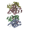
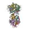
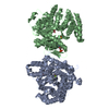
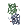
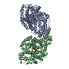
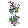
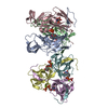
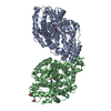
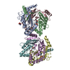
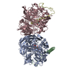
| Title | CRYSTAL STRUCTURE OF THE QUINOPROTEIN ETHANOL DEHYDROGENASE FROM PSEUDOMONAS AERUGINOSA | |||||||||
 Components Components | PROTEIN (QUINOPROTEIN ETHANOL DEHYDROGENASE) | |||||||||
 Keywords Keywords | OXIDOREDUCTASE / QUINOPROTEIN / SUPERBARREL / DEHYDROGENASE | |||||||||
| Function / homology |  Function and homology information Function and homology informationalcohol dehydrogenase (cytochrome c) / alcohol dehydrogenase (cytochrome c) activity / pyrroloquinoline quinone binding / ethanol catabolic process / outer membrane-bounded periplasmic space / heme binding / calcium ion binding / membrane Similarity search - Function | |||||||||
| Biological species |   Pseudomonas aeruginosa (bacteria) Pseudomonas aeruginosa (bacteria) | |||||||||
| Method |  X-RAY DIFFRACTION / SYNCHROTRON / Resolution: 2.6 Å X-RAY DIFFRACTION / SYNCHROTRON / Resolution: 2.6 Å | |||||||||
 Authors Authors | Keitel, T. / Diehl, A. / Knaute, T. / Stezowski, J.J. / Hohne, W. / Gorisch, H. | |||||||||
 Citation Citation | Journal: J.Mol.Biol. / Year: 2000 Title: X-ray structure of the quinoprotein ethanol dehydrogenase from Pseudomonas aeruginosa: basis of substrate specificity. Authors: Keitel, T. / Diehl, A. / Knaute, T. / Stezowski, J.J. / Hohne, W. / Gorisch, H. | |||||||||
| History |
|
- Structure visualization
| Structure viewer | Molecule: MolmilJmol/JSmol |
|---|
- Downloads & links
-Download
| PDBx/mmCIF format | 1flg.cif.gz | 241.4 KB | Display | PDBx/mmCIF format |
|---|---|---|---|---|
| PDB format | pdb1flg.ent.gz | 189.4 KB | Display | PDB format |
| PDBx/mmJSON format | 1flg.json.gz | Tree view | PDBx/mmJSON format | |
| Others |  Other downloads Other downloads |
-Validation report
| Arichive directory | https://data.pdbj.org/pub/pdb/validation_reports/fl/1flgftp://data.pdbj.org/pub/pdb/validation_reports/fl/1flg | HTTPS FTP |
|---|
-Related structure data
| Similar structure data |
|---|
-Links
 PDBj
PDBj
- Assembly
| Deposited unit | 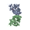
| ||||||||
|---|---|---|---|---|---|---|---|---|---|
| 1 |
| ||||||||
| Unit cell |
|
-Components
| #1: Protein | Mass: 64123.270 Da / Num. of mol.: 2 / Source method: isolated from a natural source / Source: (natural) Pseudomonas aeruginosa (bacteria) / References: UniProt: Q9Z4J7#2: Chemical | ChemComp-CA /   Mass: 40.078 Da / Num. of mol.: 4 / Source method: obtained synthetically / Formula: Ca Mass: 40.078 Da / Num. of mol.: 4 / Source method: obtained synthetically / Formula: Ca#3: Chemical |   Mass: 330.206 Da / Num. of mol.: 2 / Source method: obtained synthetically / Formula: C14H6N2O8 Mass: 330.206 Da / Num. of mol.: 2 / Source method: obtained synthetically / Formula: C14H6N2O8#4: Water | ChemComp-HOH / |  Mass: 18.015 Da / Num. of mol.: 106 / Source method: isolated from a natural source / Formula: H2O Mass: 18.015 Da / Num. of mol.: 106 / Source method: isolated from a natural source / Formula: H2OHas protein modification | Y | |
|---|
-Experimental details
-Experiment
| Experiment | Method: X-RAY DIFFRACTION / Number of used crystals: 1 |
|---|
- Sample preparation
| Crystal | Density Matthews: 2.5 Å3/Da / Density % sol: 50.7 % | ||||||||||||||||||||||||||||||||||||||||||||||||||||||||||||||||||||||
|---|---|---|---|---|---|---|---|---|---|---|---|---|---|---|---|---|---|---|---|---|---|---|---|---|---|---|---|---|---|---|---|---|---|---|---|---|---|---|---|---|---|---|---|---|---|---|---|---|---|---|---|---|---|---|---|---|---|---|---|---|---|---|---|---|---|---|---|---|---|---|---|
| Crystal grow | Temperature: 293 K / Method: vapor diffusion, hanging drop / pH: 8 Details: 10% PEG1500, 50 MM CALCIUM CHLORIDE, 4.5 MM GLYCINE/NAOH PH 8, pH 8.00, VAPOR DIFFUSION, HANGING DROP, temperature 293.0K | ||||||||||||||||||||||||||||||||||||||||||||||||||||||||||||||||||||||
| Crystal grow | *PLUS pH: 8 / Details: Stezowski, J.J., (1989) J. Mol. Biol., 205, 617. | ||||||||||||||||||||||||||||||||||||||||||||||||||||||||||||||||||||||
| Components of the solutions | *PLUS
|
-Data collection
| Diffraction | Mean temperature: 293 K |
|---|---|
| Diffraction source | Source: SYNCHROTRON / Site: EMBL/DESY, HAMBURG  / Beamline: X31 / Wavelength: 0.97 / Beamline: X31 / Wavelength: 0.97 |
| Detector | Type: MARRESEARCH / Detector: IMAGE PLATE / Date: Jun 29, 1989 |
| Radiation | Protocol: SINGLE WAVELENGTH / Monochromatic (M) / Laue (L): M / Scattering type: x-ray |
| Radiation wavelength | Wavelength: 0.97 Å / Relative weight: 1 |
| Reflection | Resolution: 2.52→25.7 Å / Num. obs: 41300 / % possible obs: 94.2 % / Observed criterion σ(I): 1 / Redundancy: 2.7 % / Biso Wilson estimate: 28.2 Å2 / Rmerge(I) obs: 0.12 / Net I/σ(I): 15 |
| Reflection shell | Resolution: 2.6→2.7 Å / Redundancy: 2.7 % / Rmerge(I) obs: 0.33 / % possible all: 98.8 |
| Reflection | *PLUS Highest resolution: 2.52 Å / Lowest resolution: 25.7 Å / Observed criterion σ(I): 1 / Redundancy: 2.7 % / Num. measured all: 109117 / Rmerge(I) obs: 0.126 / Biso Wilson estimate: 28.2 Å2 |
| Reflection shell | *PLUS Highest resolution: 2.6 Å / Lowest resolution: 2.7 Å / Rmerge(I) obs: 0.36 |
- Processing
| Software |
| |||||||||||||||||||||
|---|---|---|---|---|---|---|---|---|---|---|---|---|---|---|---|---|---|---|---|---|---|---|
| Refinement | Resolution: 2.6→12.5 Å / σ(F): 1 / σ(I): 1 / Details: MAXIMUM LIKELIHOOD
| |||||||||||||||||||||
| Refinement step | Cycle: LAST / Resolution: 2.6→12.5 Å
| |||||||||||||||||||||
| Refine LS restraints | Type: p_bond_d / Dev ideal: 0.013 | |||||||||||||||||||||
| Software | *PLUS Name: REFMAC / Classification: refinement | |||||||||||||||||||||
| Refinement | *PLUS Rfactor obs: 0.192 | |||||||||||||||||||||
| Solvent computation | *PLUS | |||||||||||||||||||||
| Displacement parameters | *PLUS Biso mean: 28.2 Å2 | |||||||||||||||||||||
| Refine LS restraints | *PLUS
|
