 Movie
Movie Controller

[English] 日本語
 Yorodumi
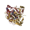
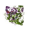
- PDB-5ojj: Crystal structure of the Zn-bound ubiquitin-conjugating enzyme Ube2T -
+ Open data

- Basic information
| Entry | Database: PDB / ID: 5ojj | ||||||
|---|---|---|---|---|---|---|---|
| Title | Crystal structure of the Zn-bound ubiquitin-conjugating enzyme Ube2T | ||||||
 Components Components | Ubiquitin-conjugating enzyme E2 T | ||||||
 Keywords Keywords | LIGASE / Ubiquitin conjugating enzyme / zinc / domain swap / oligomer | ||||||
| Function / homology |  Function and homology information Function and homology informationprotein K29-linked ubiquitination / protein K11-linked ubiquitination / protein K6-linked ubiquitination / protein K27-linked ubiquitination / E2 ubiquitin-conjugating enzyme / ubiquitin conjugating enzyme activity / protein K63-linked ubiquitination / protein monoubiquitination / protein autoubiquitination / protein K48-linked ubiquitination ...protein K29-linked ubiquitination / protein K11-linked ubiquitination / protein K6-linked ubiquitination / protein K27-linked ubiquitination / E2 ubiquitin-conjugating enzyme / ubiquitin conjugating enzyme activity / protein K63-linked ubiquitination / protein monoubiquitination / protein autoubiquitination / protein K48-linked ubiquitination / Synthesis of active ubiquitin: roles of E1 and E2 enzymes / Fanconi Anemia Pathway / protein polyubiquitination / ubiquitin-protein transferase activity / DNA repair / chromatin binding / DNA damage response / ubiquitin protein ligase binding / nucleolus / nucleoplasm / ATP binding / nucleus Similarity search - Function | ||||||
| Biological species |  Homo sapiens (human) Homo sapiens (human) | ||||||
| Method |  X-RAY DIFFRACTION / SYNCHROTRON / MOLECULAR REPLACEMENT / Resolution: 1.85 Å X-RAY DIFFRACTION / SYNCHROTRON / MOLECULAR REPLACEMENT / Resolution: 1.85 Å | ||||||
 Authors Authors | Morreale, F.E. / Testa, A. / Chaugule, V.K. / Bortoluzzi, A. / Ciulli, A. / Walden, H. | ||||||
 Citation Citation | Journal: J. Med. Chem. / Year: 2017 Title: Mind the Metal: A Fragment Library-Derived Zinc Impurity Binds the E2 Ubiquitin-Conjugating Enzyme Ube2T and Induces Structural Rearrangements. Authors: Morreale, F.E. / Testa, A. / Chaugule, V.K. / Bortoluzzi, A. / Ciulli, A. / Walden, H. | ||||||
| History |
|
- Structure visualization
| Structure viewer | Molecule: MolmilJmol/JSmol |
|---|
- Downloads & links
-Download
| PDBx/mmCIF format | 5ojj.cif.gz | 213.8 KB | Display | PDBx/mmCIF format |
|---|---|---|---|---|
| PDB format | pdb5ojj.ent.gz | 170.5 KB | Display | PDB format |
| PDBx/mmJSON format | 5ojj.json.gz | Tree view | PDBx/mmJSON format | |
| Others |  Other downloads Other downloads |
-Validation report
| Arichive directory | https://data.pdbj.org/pub/pdb/validation_reports/oj/5ojjftp://data.pdbj.org/pub/pdb/validation_reports/oj/5ojj | HTTPS FTP |
|---|
-Related structure data
| Related structure data | 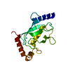 1yh2S S: Starting model for refinement |
|---|---|
| Similar structure data |


-Links
 PDBj
PDBj

- Assembly
| Deposited unit | 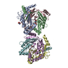
| ||||||||
|---|---|---|---|---|---|---|---|---|---|
| 1 | 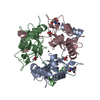
| ||||||||
| 2 | 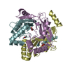
| ||||||||
| Unit cell |
|
-Components
| #1: Protein | Mass: 17686.469 Da / Num. of mol.: 6 Source method: isolated from a genetically manipulated source Source: (gene. exp.) Homo sapiens (human) / Gene: UBE2T, HSPC150, PIG50 / Production host:  References: UniProt: Q9NPD8, E2 ubiquitin-conjugating enzyme #2: Chemical | ChemComp-ACT /   Mass: 59.044 Da / Num. of mol.: 6 / Source method: obtained synthetically / Formula: C2H3O2 Mass: 59.044 Da / Num. of mol.: 6 / Source method: obtained synthetically / Formula: C2H3O2#3: Chemical | ChemComp-ZN /   Mass: 65.409 Da / Num. of mol.: 8 / Source method: obtained synthetically / Formula: Zn Mass: 65.409 Da / Num. of mol.: 8 / Source method: obtained synthetically / Formula: Zn#4: Chemical |   Mass: 122.143 Da / Num. of mol.: 2 / Source method: obtained synthetically / Formula: C4H12NO3 / Comment: pH buffer*YM Mass: 122.143 Da / Num. of mol.: 2 / Source method: obtained synthetically / Formula: C4H12NO3 / Comment: pH buffer*YM#5: Water | ChemComp-HOH / |  Mass: 18.015 Da / Num. of mol.: 978 / Source method: isolated from a natural source / Formula: H2O Mass: 18.015 Da / Num. of mol.: 978 / Source method: isolated from a natural source / Formula: H2O |
|---|
-Experimental details
-Experiment
| Experiment | Method: X-RAY DIFFRACTION / Number of used crystals: 1 |
|---|
- Sample preparation
| Crystal | Density Matthews: 2.29 Å3/Da / Density % sol: 46.19 % |
|---|---|
| Crystal grow | Temperature: 293 K / Method: vapor diffusion, sitting drop Details: 10% PEG3350, 0.2 M calcium acetate, 0.1 M Tris pH 8.5 |
-Data collection
| Diffraction | Mean temperature: 100 K |
|---|---|
| Diffraction source | Source: SYNCHROTRON / Site: Diamond  / Beamline: I04-1 / Wavelength: 0.9282 Å / Beamline: I04-1 / Wavelength: 0.9282 Å |
| Detector | Type: DECTRIS PILATUS 2M / Detector: PIXEL / Date: Sep 25, 2016 |
| Radiation | Protocol: SINGLE WAVELENGTH / Monochromatic (M) / Laue (L): M / Scattering type: x-ray |
| Radiation wavelength | Wavelength: 0.9282 Å / Relative weight: 1 |
| Reflection | Resolution: 1.85→48.75 Å / Num. obs: 81137 / % possible obs: 99.7 % / Redundancy: 3.4 % / CC1/2: 0.998 / Rmerge(I) obs: 0.038 / Net I/σ(I): 16.6 |
| Reflection shell | Resolution: 1.85→1.89 Å / Redundancy: 3.1 % / Rmerge(I) obs: 0.292 / Mean I/σ(I) obs: 3.1 / Num. unique obs: 4465 / CC1/2: 0.849 / % possible all: 99.6 |
- Processing
| Software |
| ||||||||||||||||||||||||||||||||||||||||||||||||||||||||||||||||||||||||||||||||||||||||||||||||||||||||||||||||||||||||||||||||||||||||||||||||||||||||||||||||||||||||||||||||||||||
|---|---|---|---|---|---|---|---|---|---|---|---|---|---|---|---|---|---|---|---|---|---|---|---|---|---|---|---|---|---|---|---|---|---|---|---|---|---|---|---|---|---|---|---|---|---|---|---|---|---|---|---|---|---|---|---|---|---|---|---|---|---|---|---|---|---|---|---|---|---|---|---|---|---|---|---|---|---|---|---|---|---|---|---|---|---|---|---|---|---|---|---|---|---|---|---|---|---|---|---|---|---|---|---|---|---|---|---|---|---|---|---|---|---|---|---|---|---|---|---|---|---|---|---|---|---|---|---|---|---|---|---|---|---|---|---|---|---|---|---|---|---|---|---|---|---|---|---|---|---|---|---|---|---|---|---|---|---|---|---|---|---|---|---|---|---|---|---|---|---|---|---|---|---|---|---|---|---|---|---|---|---|---|---|
| Refinement | Method to determine structure: MOLECULAR REPLACEMENT Starting model: 1YH2 Resolution: 1.85→48.75 Å / Cor.coef. Fo:Fc: 0.962 / Cor.coef. Fo:Fc free: 0.953 / SU B: 2.758 / SU ML: 0.083 / Cross valid method: THROUGHOUT / ESU R: 0.145 / ESU R Free: 0.123 / Details: HYDROGENS HAVE BEEN ADDED IN THE RIDING POSITIONS
| ||||||||||||||||||||||||||||||||||||||||||||||||||||||||||||||||||||||||||||||||||||||||||||||||||||||||||||||||||||||||||||||||||||||||||||||||||||||||||||||||||||||||||||||||||||||
| Solvent computation | Ion probe radii: 0.8 Å / Shrinkage radii: 0.8 Å / VDW probe radii: 1.2 Å | ||||||||||||||||||||||||||||||||||||||||||||||||||||||||||||||||||||||||||||||||||||||||||||||||||||||||||||||||||||||||||||||||||||||||||||||||||||||||||||||||||||||||||||||||||||||
| Displacement parameters | Biso mean: 28.966 Å2
| ||||||||||||||||||||||||||||||||||||||||||||||||||||||||||||||||||||||||||||||||||||||||||||||||||||||||||||||||||||||||||||||||||||||||||||||||||||||||||||||||||||||||||||||||||||||
| Refinement step | Cycle: 1 / Resolution: 1.85→48.75 Å
| ||||||||||||||||||||||||||||||||||||||||||||||||||||||||||||||||||||||||||||||||||||||||||||||||||||||||||||||||||||||||||||||||||||||||||||||||||||||||||||||||||||||||||||||||||||||
| Refine LS restraints |
|