| Entry | Database: PDB / ID: 4wrk
|
|---|
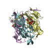
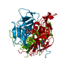
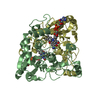
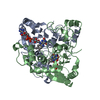
| Title | The 3D structure of D95N mutant DUTPase from phage phi11 of S. aureus reveals the molecular details for the coordination of a structural Mg(II) ion |
|---|
 Components Components | DUTPase |
|---|
 Keywords Keywords | HYDROLASE / Magnesium coordination mutant / jelly-roll |
|---|
| Function / homology |  Function and homology information Function and homology information
dUTP catabolic process / dUMP biosynthetic process / dUTP diphosphatase / dUTP diphosphatase activity / magnesium ion bindingSimilarity search - Function Deoxyuridine triphosphate nucleotidohydrolase / Deoxyuridine triphosphatase (dUTPase) / Deoxyuridine 5'-Triphosphate Nucleotidohydrolase; Chain A / dUTPase-like / dUTPase / dUTPase, trimeric / dUTPase-like superfamily / Distorted Sandwich / Mainly BetaSimilarity search - Domain/homology |
|---|
| Biological species |  Staphylococcus phage phi11 (virus) Staphylococcus phage phi11 (virus) |
|---|
| Method |  X-RAY DIFFRACTION / MOLECULAR REPLACEMENT / Resolution: 2.9 Å X-RAY DIFFRACTION / MOLECULAR REPLACEMENT / Resolution: 2.9 Å |
|---|
 Authors Authors | Bendes, A.A. / Leveles, I. / Vertessy, B.G. |
|---|
| Funding support |  Hungary, 6items Hungary, 6items | Organization | Grant number | Country |
|---|
| ungarian Scientific Research Fund OTKA | NK 84008 | Hungary | | ungarian Scientific Research Fund OTKA | K109486 | Hungary | | Baross Program of the New Hungary Development Plan | 3DSTRUCT, MFB-00266/2010 REG-KM-09-1-2009-005 | Hungary | | Hungarian Academy of Sciences | TTK IF-28/201 | Hungary | | Hungarian Academy of Sciences | MedinProt program | Hungary | | European Union | 283570 | |
|
|---|
 Citation Citation | Journal: To Be Published
Title: The 3D structure of D95N mutant DUTPase from phage phi11 of S. aureus reveals the molecular details for the coordination of a structural Mg(II) ion
Authors: Bendes, A.A. / Leveles, I. / Vertessy, B.G. |
|---|
| History | | Deposition | Oct 24, 2014 | Deposition site: RCSB / Processing site: PDBE |
|---|
| Revision 1.0 | Dec 23, 2015 | Provider: repository / Type: Initial release |
|---|
| Revision 2.0 | Jan 10, 2024 | Group: Atomic model / Author supporting evidence ...Atomic model / Author supporting evidence / Data collection / Database references / Derived calculations / Refinement description
Category: atom_site / chem_comp_atom ...atom_site / chem_comp_atom / chem_comp_bond / database_2 / pdbx_audit_support / pdbx_initial_refinement_model / pdbx_nonpoly_scheme / pdbx_struct_conn_angle / struct_conn / struct_site_gen
Item: _atom_site.B_iso_or_equiv / _atom_site.Cartn_x ..._atom_site.B_iso_or_equiv / _atom_site.Cartn_x / _atom_site.Cartn_y / _atom_site.Cartn_z / _atom_site.occupancy / _database_2.pdbx_DOI / _database_2.pdbx_database_accession / _pdbx_audit_support.funding_organization / _pdbx_nonpoly_scheme.auth_seq_num / _pdbx_struct_conn_angle.ptnr3_auth_seq_id / _struct_conn.ptnr2_auth_seq_id / _struct_site_gen.auth_seq_id |
|---|
|
|---|
 Movie
Movie Controller

 Yorodumi
 Open data
 Basic information
 Structure visualization
 Downloads & links
 Other downloads

 PDBj
PDBj
 Assembly


 Mass: 467.157 Da / Num. of mol.: 6 / Source method: obtained synthetically / Formula: C9H16N3O13P3
 Mass: 24.305 Da / Num. of mol.: 6 / Source method: obtained synthetically / Formula: Mg
 Mass: 18.015 Da / Num. of mol.: 101 / Source method: isolated from a natural source / Formula: H2O
 Sample preparation
 Processing