 Movie
Movie Controller


+ Open data

- Basic information
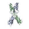
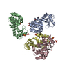
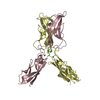
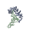
| Entry | Database: PDB / ID: 1ff5 | ||||||
|---|---|---|---|---|---|---|---|
| Title | STRUCTURE OF E-CADHERIN DOUBLE DOMAIN | ||||||
 Components Components | EPITHELIAL CADHERIN | ||||||
 Keywords Keywords | CELL ADHESION / E-cadherin / Ca-binding | ||||||
| Function / homology |  Function and homology information Function and homology informationuterine epithelium development / Apoptotic cleavage of cell adhesion proteins / Regulation of CDH1 posttranslational processing and trafficking to plasma membrane / desmosome assembly / salivary gland cavitation / regulation of branching involved in salivary gland morphogenesis / Adherens junctions interactions / Degradation of the extracellular matrix / RHO GTPases activate IQGAPs / Integrin cell surface interactions ...uterine epithelium development / Apoptotic cleavage of cell adhesion proteins / Regulation of CDH1 posttranslational processing and trafficking to plasma membrane / desmosome assembly / salivary gland cavitation / regulation of branching involved in salivary gland morphogenesis / Adherens junctions interactions / Degradation of the extracellular matrix / RHO GTPases activate IQGAPs / Integrin cell surface interactions / Regulation of CDH1 Function / Degradation of CDH1 / gamma-catenin binding / desmosome / positive regulation of cell-cell adhesion / lateral loop / regulation of protein localization to cell surface / negative regulation of axon extension / cell-cell adhesion mediator activity / alpha-catenin binding / cellular response to indole-3-methanol / trophectodermal cell differentiation / calcium-dependent cell-cell adhesion / intestinal epithelial cell development / bicellular tight junction assembly / Schmidt-Lanterman incisure / regulation of protein catabolic process at postsynapse, modulating synaptic transmission / flotillin complex / cell-cell adhesion mediated by cadherin / adherens junction organization / epithelial cell morphogenesis / catenin complex / regulation of neuron migration / node of Ranvier / negative regulation of protein processing / cell-cell junction assembly / GTPase activating protein binding / negative regulation of cell-cell adhesion / ankyrin binding / cellular response to lithium ion / apical junction complex / homophilic cell-cell adhesion / decidualization / microvillus / establishment of skin barrier / cochlea development / lateral plasma membrane / negative regulation of protein localization to plasma membrane / positive regulation of protein localization / cytoskeletal protein binding / synapse assembly / embryo implantation / cell adhesion molecule binding / protein tyrosine kinase binding / axon terminus / negative regulation of cell migration / cell periphery / protein localization to plasma membrane / adherens junction / trans-Golgi network / cellular response to amino acid stimulus / negative regulation of canonical Wnt signaling pathway / sensory perception of sound / cell-cell adhesion / positive regulation of protein import into nucleus / beta-catenin binding / cell morphogenesis / negative regulation of epithelial cell proliferation / cytoplasmic side of plasma membrane / apical part of cell / cell-cell junction / cell junction / cell migration / regulation of protein localization / lamellipodium / actin cytoskeleton / regulation of gene expression / actin cytoskeleton organization / protein phosphatase binding / molecular adaptor activity / in utero embryonic development / basolateral plasma membrane / endosome / postsynapse / cadherin binding / protein domain specific binding / axon / calcium ion binding / positive regulation of DNA-templated transcription / perinuclear region of cytoplasm / glutamatergic synapse / cell surface / membrane / identical protein binding / plasma membrane / cytoplasm Similarity search - Function | ||||||
| Biological species |  | ||||||
| Method |  X-RAY DIFFRACTION / Resolution: 2.93 Å X-RAY DIFFRACTION / Resolution: 2.93 Å | ||||||
 Authors Authors | Pertz, O. / Bozic, D. / Koch, A.W. / Fauser, C. / Brancaccio, A. / Engel, J. | ||||||
 Citation Citation | Journal: EMBO J. / Year: 1999 Title: A new crystal structure, Ca2+ dependence and mutational analysis reveal molecular details of E-cadherin homoassociation. Authors: Pertz, O. / Bozic, D. / Koch, A.W. / Fauser, C. / Brancaccio, A. / Engel, J. | ||||||
| History |
|
- Structure visualization
| Structure viewer | Molecule: MolmilJmol/JSmol |
|---|
- Downloads & links
-Download
| PDBx/mmCIF format | 1ff5.cif.gz | 103.4 KB | Display | PDBx/mmCIF format |
|---|---|---|---|---|
| PDB format | pdb1ff5.ent.gz | 78.8 KB | Display | PDB format |
| PDBx/mmJSON format | 1ff5.json.gz | Tree view | PDBx/mmJSON format | |
| Others |  Other downloads Other downloads |
-Validation report
| Arichive directory | https://data.pdbj.org/pub/pdb/validation_reports/ff/1ff5ftp://data.pdbj.org/pub/pdb/validation_reports/ff/1ff5 | HTTPS FTP |
|---|
-Related structure data
| Related structure data | |
|---|---|
| Similar structure data |


-Links
 PDBj
PDBj
- Assembly
| Deposited unit | 
| ||||||||
|---|---|---|---|---|---|---|---|---|---|
| 1 |
| ||||||||
| Unit cell |
|
-Components
| #1: Protein | Mass: 23919.590 Da / Num. of mol.: 2 / Fragment: DOUBLE DOMAIN Source method: isolated from a genetically manipulated source Source: (gene. exp.)  #2: Chemical | ChemComp-CA /   Mass: 40.078 Da / Num. of mol.: 6 / Source method: obtained synthetically / Formula: Ca Mass: 40.078 Da / Num. of mol.: 6 / Source method: obtained synthetically / Formula: Ca#3: Water | ChemComp-HOH / |  Mass: 18.015 Da / Num. of mol.: 265 / Source method: isolated from a natural source / Formula: H2O Mass: 18.015 Da / Num. of mol.: 265 / Source method: isolated from a natural source / Formula: H2O |
|---|
-Experimental details
-Experiment
| Experiment | Method: X-RAY DIFFRACTION / Number of used crystals: 1 |
|---|
- Sample preparation
| Crystal | Density Matthews: 6.92 Å3/Da | ||||||||||||||||||||||||||||||||||||||||||||||||||||||
|---|---|---|---|---|---|---|---|---|---|---|---|---|---|---|---|---|---|---|---|---|---|---|---|---|---|---|---|---|---|---|---|---|---|---|---|---|---|---|---|---|---|---|---|---|---|---|---|---|---|---|---|---|---|---|---|
| Crystal grow | Temperature: 298 K / Method: vapor diffusion, hanging drop / pH: 8.5 Details: 50 mM ammonium sulfate, 50 mM NaOAc, 30% PEG 8000, pH 8.5, Tris-HCl, VAPOR DIFFUSION, HANGING DROP, temperature 298.0K | ||||||||||||||||||||||||||||||||||||||||||||||||||||||
| Crystal | *PLUS Density % sol: 58 % | ||||||||||||||||||||||||||||||||||||||||||||||||||||||
| Crystal grow | *PLUS pH: 7.4 | ||||||||||||||||||||||||||||||||||||||||||||||||||||||
| Components of the solutions | *PLUS
|
-Data collection
| Diffraction | Mean temperature: 298 K |
|---|---|
| Diffraction source | Source: ROTATING ANODE / Type: ELLIOTT GX-20 / Wavelength: 1.5418 |
| Detector | Type: MARRESEARCH / Detector: AREA DETECTOR |
| Radiation | Protocol: SINGLE WAVELENGTH / Monochromatic (M) / Laue (L): M / Scattering type: x-ray |
| Radiation wavelength | Wavelength: 1.5418 Å / Relative weight: 1 |
| Reflection | Resolution: 2.93→20 Å / Num. all: 28520 / Num. obs: 28520 / % possible obs: 95.5 % / Rmerge(I) obs: 0.133 |
| Reflection | *PLUS Num. obs: 13451 / Num. measured all: 28520 |
| Reflection shell | *PLUS Highest resolution: 2.93 Å / Lowest resolution: 3 Å / % possible obs: 81.5 % |
- Processing
| Software |
| ||||||||||||||||||
|---|---|---|---|---|---|---|---|---|---|---|---|---|---|---|---|---|---|---|---|
| Refinement | Resolution: 2.93→20 Å / Rfactor all: 0.1986 / σ(F): 0 / Stereochemistry target values: Engh & Huber | ||||||||||||||||||
| Refinement step | Cycle: LAST / Resolution: 2.93→20 Å
| ||||||||||||||||||
| Refine LS restraints |
| ||||||||||||||||||
| Software | *PLUS Name: X-PLOR / Version: 3.1 / Classification: refinement | ||||||||||||||||||
| Refinement | *PLUS Lowest resolution: 20 Å / σ(F): 0 | ||||||||||||||||||
| Solvent computation | *PLUS | ||||||||||||||||||
| Displacement parameters | *PLUS | ||||||||||||||||||
| Refine LS restraints | *PLUS Type: x_bond_d / Dev ideal: 0.01 |
