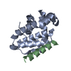
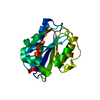
- PDB-1shu: Crystal Structure of the von Willebrand factor A domain of human ... -
+
Open data
ID or keywords:
Loading...
-
Basic information
Entry
Database: PDB / ID: 1shu
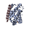
Title
Crystal Structure of the von Willebrand factor A domain of human capillary morphogenesis protein 2: an anthrax toxin receptor
Components
Anthrax toxin receptor 2
Keywords
MEMBRANE PROTEIN / alpha/beta Rossmann fold
Function / homology
Function and homology information
collagen fibril organization / uterus development / Uptake and function of anthrax toxins / single fertilization / transmembrane signaling receptor activity / endosome membrane / external side of plasma membrane / endoplasmic reticulum membrane / cell surface / extracellular region ...collagen fibril organization / uterus development / Uptake and function of anthrax toxins / single fertilization / transmembrane signaling receptor activity / endosome membrane / external side of plasma membrane / endoplasmic reticulum membrane / cell surface / extracellular region / metal ion binding / plasma membrane Similarity search - Function
Anthrax toxin receptor, C-terminal / Anthrax toxin receptor, extracellular domain / Anthrax toxin receptor / Anthrax receptor C-terminus region / Anthrax receptor extracellular domain / von Willebrand factor, type A domain / von Willebrand factor type A domain / VWFA domain profile. / von Willebrand factor (vWF) type A domain / von Willebrand factor, type A ...Anthrax toxin receptor, C-terminal / Anthrax toxin receptor, extracellular domain / Anthrax toxin receptor / Anthrax receptor C-terminus region / Anthrax receptor extracellular domain / von Willebrand factor, type A domain / von Willebrand factor type A domain / VWFA domain profile. / von Willebrand factor (vWF) type A domain / von Willebrand factor, type A / von Willebrand factor A-like domain superfamily / Rossmann fold / 3-Layer(aba) Sandwich / Alpha Beta Similarity search - Domain/homology
In the structure databanks used in Yorodumi, some data are registered as the other names, "COVID-19 virus" and "2019-nCoV". Here are the details of the virus and the list of structure data.
Jan 31, 2019. EMDB accession codes are about to change! (news from PDBe EMDB page)
EMDB accession codes are about to change! (news from PDBe EMDB page)
The allocation of 4 digits for EMDB accession codes will soon come to an end. Whilst these codes will remain in use, new EMDB accession codes will include an additional digit and will expand incrementally as the available range of codes is exhausted. The current 4-digit format prefixed with “EMD-” (i.e. EMD-XXXX) will advance to a 5-digit format (i.e. EMD-XXXXX), and so on. It is currently estimated that the 4-digit codes will be depleted around Spring 2019, at which point the 5-digit format will come into force.
The EM Navigator/Yorodumi systems omit the EMD- prefix.
Related info.:Q: What is EMD? / ID/Accession-code notation in Yorodumi/EM Navigator
Yorodumi is a browser for structure data from EMDB, PDB, SASBDB, etc.
This page is also the successor to EM Navigator detail page, and also detail information page/front-end page for Omokage search.
The word "yorodu" (or yorozu) is an old Japanese word meaning "ten thousand". "mi" (miru) is to see.
Related info.:EMDB / PDB / SASBDB / Comparison of 3 databanks / Yorodumi Search / Aug 31, 2016. New EM Navigator & Yorodumi / Yorodumi Papers / Jmol/JSmol / Function and homology information / Changes in new EM Navigator and Yorodumi
 Movie
Movie Controller

 Yorodumi
 Open data
 Basic information
 Components
 Keywords
 Function and homology information
 Homo sapiens (human)
 X-RAY DIFFRACTION /
  Authors
 Citation
 Structure visualization
 Downloads & links
 Other downloads


 PDBj
PDBj


 Assembly


 Mass: 24.305 Da / Num. of mol.: 1 / Source method: obtained synthetically / Formula: Mg
 Mass: 18.015 Da / Num. of mol.: 154 / Source method: isolated from a natural source / Formula: H2O
 Sample preparation
 / Beamline: F1 / Wavelength: 0.9186 Å
 Processing