 Movie
Movie Controller


+ Open data

- Basic information
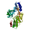
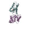
| Entry | Database: PDB / ID: 1fc6 | ||||||
|---|---|---|---|---|---|---|---|
| Title | PHOTOSYSTEM II D1 C-TERMINAL PROCESSING PROTEASE | ||||||
 Components Components | PHOTOSYSTEM II D1 PROTEASE | ||||||
 Keywords Keywords | HYDROLASE / D1 C-terminal processing protease / serine protease / serine-lysine catalytic dyad / PDZ domain / photosystem II / photosynthesis / x-ray crystal structure | ||||||
| Function / homology |  Function and homology information Function and homology informationC-terminal processing peptidase / chloroplast thylakoid lumen / serine-type endopeptidase activity / proteolysis Similarity search - Function | ||||||
| Biological species |  Scenedesmus obliquus (plant) Scenedesmus obliquus (plant) | ||||||
| Method |  X-RAY DIFFRACTION / SYNCHROTRON / MAD, treating MAD data as a special case of MIR / Resolution: 1.8 Å X-RAY DIFFRACTION / SYNCHROTRON / MAD, treating MAD data as a special case of MIR / Resolution: 1.8 Å | ||||||
 Authors Authors | Liao, D.I. / Qian, J. / Chisholm, D.A. / Jordan, D.B. / Diner, B.A. | ||||||
 Citation Citation | Journal: Nat.Struct.Biol. / Year: 2000 Title: Crystal structures of the photosystem II D1 C-terminal processing protease. Authors: Liao, D.I. / Qian, J. / Chisholm, D.A. / Jordan, D.B. / Diner, B.A. | ||||||
| History |
|
- Structure visualization
| Structure viewer | Molecule: MolmilJmol/JSmol |
|---|
- Downloads & links
-Download
| PDBx/mmCIF format | 1fc6.cif.gz | 92.5 KB | Display | PDBx/mmCIF format |
|---|---|---|---|---|
| PDB format | pdb1fc6.ent.gz | 68.7 KB | Display | PDB format |
| PDBx/mmJSON format | 1fc6.json.gz | Tree view | PDBx/mmJSON format | |
| Others |  Other downloads Other downloads |
-Validation report
| Arichive directory | https://data.pdbj.org/pub/pdb/validation_reports/fc/1fc6ftp://data.pdbj.org/pub/pdb/validation_reports/fc/1fc6 | HTTPS FTP |
|---|
-Related structure data


-Links
 PDBj
PDBj
- Assembly
| Deposited unit | 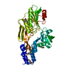
| ||||||||
|---|---|---|---|---|---|---|---|---|---|
| 1 |
| ||||||||
| Unit cell |
| ||||||||
| Components on special symmetry positions |
|
-Components
| #1: Protein | Mass: 41019.566 Da / Num. of mol.: 1 / Fragment: RESIDUES 77-464 / Mutation: L132M, L210M Source method: isolated from a genetically manipulated source Source: (gene. exp.) Scenedesmus obliquus (plant) / Plasmid: PET-32(A)-D1P(+AM) / Production host:  |
|---|---|
| #2: Water | ChemComp-HOH /  Mass: 18.015 Da / Num. of mol.: 352 / Source method: isolated from a natural source / Formula: H2O Mass: 18.015 Da / Num. of mol.: 352 / Source method: isolated from a natural source / Formula: H2O |
| Has protein modification | Y |
-Experimental details
-Experiment
| Experiment | Method: X-RAY DIFFRACTION / Number of used crystals: 1 |
|---|
- Sample preparation
| Crystal | Density Matthews: 2.36 Å3/Da / Density % sol: 47.86 % | |||||||||||||||||||||||||||||||||||
|---|---|---|---|---|---|---|---|---|---|---|---|---|---|---|---|---|---|---|---|---|---|---|---|---|---|---|---|---|---|---|---|---|---|---|---|---|
| Crystal grow | Temperature: 298 K / Method: vapor diffusion, hanging drop / pH: 7.8 Details: PEG 4000, isopropanol, HEPES, pH 7.8, VAPOR DIFFUSION, HANGING DROP, temperature 298.0K | |||||||||||||||||||||||||||||||||||
| Crystal grow | *PLUS pH: 7.5 | |||||||||||||||||||||||||||||||||||
| Components of the solutions | *PLUS
|
-Data collection
| Diffraction | Mean temperature: 102 K |
|---|---|
| Diffraction source | Source: SYNCHROTRON / Site: APS  / Beamline: 5ID-B / Wavelength: 0.9946 / Beamline: 5ID-B / Wavelength: 0.9946 |
| Detector | Type: MARRESEARCH / Detector: CCD / Date: Nov 20, 1998 |
| Radiation | Protocol: MAD / Monochromatic (M) / Laue (L): M / Scattering type: x-ray |
| Radiation wavelength | Wavelength: 0.9946 Å / Relative weight: 1 |
| Reflection | Resolution: 1.8→30 Å / Num. all: 258532 / Num. obs: 35489 / % possible obs: 99.8 % / Observed criterion σ(I): -3 / Redundancy: 7.28 % / Rmerge(I) obs: 0.056 / Net I/σ(I): 18 |
| Reflection shell | Resolution: 1.8→1.83 Å / Redundancy: 6.5 % / Rmerge(I) obs: 0.394 / Num. unique all: 1737 / % possible all: 99 |
| Reflection | *PLUS Num. measured all: 258532 |
- Processing
| Software |
| ||||||||||||||||||||
|---|---|---|---|---|---|---|---|---|---|---|---|---|---|---|---|---|---|---|---|---|---|
| Refinement | Method to determine structure: MAD, treating MAD data as a special case of MIR Resolution: 1.8→30 Å / σ(F): 2 Stereochemistry target values: TNT library, Bond lengths 0.02 bond angles 3.00
| ||||||||||||||||||||
| Solvent computation | Solvent model: Method implemented in the refinement program TNT Bsol: 248.14 Å2 / ksol: 0.86085 e/Å3 | ||||||||||||||||||||
| Refinement step | Cycle: LAST / Resolution: 1.8→30 Å
| ||||||||||||||||||||
| Refine LS restraints |
| ||||||||||||||||||||
| Software | *PLUS Name: TNT / Version: 5E / Classification: refinement | ||||||||||||||||||||
| Refinement | *PLUS Highest resolution: 1.8 Å / σ(F): 2 / % reflection Rfree: 10 % / Rfactor obs: 0.168 | ||||||||||||||||||||
| Solvent computation | *PLUS | ||||||||||||||||||||
| Displacement parameters | *PLUS | ||||||||||||||||||||
| Refine LS restraints | *PLUS Type: t_angle_deg / Dev ideal: 2.4 |
