 Movie
Movie Controller


+ Open data

- Basic information
| Entry | Database: PDB / ID: 1f8w | ||||||
|---|---|---|---|---|---|---|---|
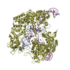
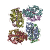
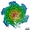
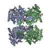
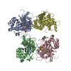
| Title | CRYSTAL STRUCTURE OF NADH PEROXIDASE MUTANT: R303M | ||||||
 Components Components | NADH PEROXIDASE | ||||||
 Keywords Keywords | OXIDOREDUCTASE / Interface / FAD / NAD-binding domains | ||||||
| Function / homology |  Function and homology information Function and homology information | ||||||
| Biological species |   Enterococcus faecalis (bacteria) Enterococcus faecalis (bacteria) | ||||||
| Method |  X-RAY DIFFRACTION / SYNCHROTRON / Resolution: 2.45 Å X-RAY DIFFRACTION / SYNCHROTRON / Resolution: 2.45 Å | ||||||
 Authors Authors | Yeh, J.I. / Claiborne, A. / Hol, W.G.J. | ||||||
 Citation Citation | Journal: Biochemistry / Year: 2000 Title: Analysis of the kinetic and redox properties of the NADH peroxidase R303M mutant: correlation with the crystal structure. Authors: Crane III, E.J. / Yeh, J.I. / Luba, J. / Claiborne, A. #1: Journal: Biochemistry / Year: 1996Title: Structure of the Native Cysteine-sulfenic Acid Redox Center of Enterococcal NADH Peroxidase Refined at 2.8 A Resolution Authors: Yeh, J.I. / Claiborne, A. / Hol, W.G. | ||||||
| History |
|
- Structure visualization
| Structure viewer | Molecule: MolmilJmol/JSmol |
|---|
- Downloads & links
-Download
| PDBx/mmCIF format | 1f8w.cif.gz | 98.7 KB | Display | PDBx/mmCIF format |
|---|---|---|---|---|
| PDB format | pdb1f8w.ent.gz | 80.6 KB | Display | PDB format |
| PDBx/mmJSON format | 1f8w.json.gz | Tree view | PDBx/mmJSON format | |
| Others |  Other downloads Other downloads |
-Validation report
| Arichive directory | https://data.pdbj.org/pub/pdb/validation_reports/f8/1f8wftp://data.pdbj.org/pub/pdb/validation_reports/f8/1f8w | HTTPS FTP |
|---|
-Related structure data
| Related structure data | |
|---|---|
| Similar structure data |


-Links
 PDBj
PDBj


- Assembly
| Deposited unit | 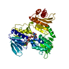
| ||||||||
|---|---|---|---|---|---|---|---|---|---|
| 1 | 
| ||||||||
| 2 | 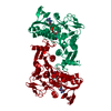
| ||||||||
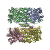
| Unit cell |
| ||||||||
| Details | FUNCTIONAL MOLECULE IS A HOMOTETRAMER |
-Components
| #1: Protein | Mass: 49593.230 Da / Num. of mol.: 1 / Mutation: ARG 303 TO MET 303 Source method: isolated from a genetically manipulated source Source: (gene. exp.) Enterococcus faecalis (bacteria) / Plasmid: PR303M / Production host: |
|---|---|
| #2: Chemical | ChemComp-FAD /   Mass: 785.550 Da / Num. of mol.: 1 / Source method: obtained synthetically / Formula: C27H33N9O15P2 / Comment: FAD*YM Mass: 785.550 Da / Num. of mol.: 1 / Source method: obtained synthetically / Formula: C27H33N9O15P2 / Comment: FAD*YM |
| #3: Water | ChemComp-HOH /  Mass: 18.015 Da / Num. of mol.: 131 / Source method: isolated from a natural source / Formula: H2O Mass: 18.015 Da / Num. of mol.: 131 / Source method: isolated from a natural source / Formula: H2O |
-Experimental details
-Experiment
| Experiment | Method: X-RAY DIFFRACTION / Number of used crystals: 1 |
|---|
- Sample preparation
| Crystal | Density Matthews: 2.8 Å3/Da | |||||||||||||||||||||||||
|---|---|---|---|---|---|---|---|---|---|---|---|---|---|---|---|---|---|---|---|---|---|---|---|---|---|---|
| Crystal grow | Temperature: 296 K / Method: vapor diffusion, hanging drop / pH: 7.2 Details: 2% PEG 400, 2.0 M (NH4)2SO4, 100 mM Na Hepes, pH 7.2, VAPOR DIFFUSION, HANGING DROP, temperature 296K | |||||||||||||||||||||||||
| Crystal grow | *PLUS Method: vapor diffusion | |||||||||||||||||||||||||
| Components of the solutions | *PLUS
|
-Data collection
| Diffraction | Mean temperature: 110 K |
|---|---|
| Diffraction source | Source: SYNCHROTRON / Site: SSRL  / Beamline: BL7-1 / Wavelength: 1.08 / Beamline: BL7-1 / Wavelength: 1.08 |
| Detector | Type: MARRESEARCH / Detector: IMAGE PLATE / Date: Aug 8, 1996 |
| Radiation | Protocol: SINGLE WAVELENGTH / Monochromatic (M) / Laue (L): M / Scattering type: x-ray |
| Radiation wavelength | Wavelength: 1.08 Å / Relative weight: 1 |
| Reflection | Resolution: 2.45→99 Å / Num. all: 42662 / Num. obs: 36615 / % possible obs: 85.8 % / Observed criterion σ(F): 2 / Observed criterion σ(I): 2 / Redundancy: 5.4 % / Biso Wilson estimate: 10.2 Å2 / Rmerge(I) obs: 0.055 / Net I/σ(I): 9.5 |
| Reflection shell | Highest resolution: 2.45 Å / Redundancy: 3.5 % / Rmerge(I) obs: 0.363 / Mean I/σ(I) obs: 2.6 / Rsym value: 0.39 / % possible all: 71.1 |
| Reflection | *PLUS Num. measured all: 60231 / Rmerge(I) obs: 0.048 |
| Reflection shell | *PLUS Lowest resolution: 2.56 Å / % possible obs: 70.9 % |
- Processing
| Software |
| |||||||||||||||||||||||||
|---|---|---|---|---|---|---|---|---|---|---|---|---|---|---|---|---|---|---|---|---|---|---|---|---|---|---|
| Refinement | Resolution: 2.45→50 Å / Cross valid method: X-PLOR FreeR / σ(F): 2 / σ(I): 2 / Stereochemistry target values: Engh & Huber
| |||||||||||||||||||||||||
| Refine analyze | Luzzati sigma a obs: 0.26 Å | |||||||||||||||||||||||||
| Refinement step | Cycle: LAST / Resolution: 2.45→50 Å
| |||||||||||||||||||||||||
| Refine LS restraints |
| |||||||||||||||||||||||||
| Software | *PLUS Name: X-PLOR / Version: 3.1 / Classification: refinement | |||||||||||||||||||||||||
| Refinement | *PLUS Lowest resolution: 50 Å / σ(F): 2 / % reflection Rfree: 5 % / Rfactor obs: 0.196 | |||||||||||||||||||||||||
| Solvent computation | *PLUS | |||||||||||||||||||||||||
| Displacement parameters | *PLUS Biso mean: 39.1 Å2 | |||||||||||||||||||||||||
| Refine LS restraints | *PLUS
|
