 Movie
Movie Controller

[English] 日本語
 Yorodumi
- PDB-1f8i: CRYSTAL STRUCTURE OF ISOCITRATE LYASE:NITROPROPIONATE:GLYOXYLATE ... -
+ Open data

- Basic information
| Entry | Database: PDB / ID: 1f8i | ||||||
|---|---|---|---|---|---|---|---|
| Title | CRYSTAL STRUCTURE OF ISOCITRATE LYASE:NITROPROPIONATE:GLYOXYLATE COMPLEX FROM MYCOBACTERIUM TUBERCULOSIS | ||||||
 Components Components | ISOCITRATE LYASE | ||||||
 Keywords Keywords | LYASE / AlPHA-BETA Barrel / Swapped Helices / closed conformation / Structural Genomics / PSI / Protein Structure Initiative / TB Structural Genomics Consortium / TBSGC | ||||||
| Function / homology |  Function and homology information Function and homology informationmethylisocitrate lyase / isocitrate lyase / isocitrate lyase activity / methylisocitrate lyase activity / isocitrate metabolic process / zymogen binding / glyoxylate cycle / tricarboxylic acid cycle / cellular response to hypoxia / extracellular region ...methylisocitrate lyase / isocitrate lyase / isocitrate lyase activity / methylisocitrate lyase activity / isocitrate metabolic process / zymogen binding / glyoxylate cycle / tricarboxylic acid cycle / cellular response to hypoxia / extracellular region / metal ion binding / plasma membrane / cytosol Similarity search - Function | ||||||
| Biological species |  Mycobacterium tuberculosis H37Rv (bacteria) Mycobacterium tuberculosis H37Rv (bacteria) | ||||||
| Method |  X-RAY DIFFRACTION / SYNCHROTRON / Resolution: 2.25 Å X-RAY DIFFRACTION / SYNCHROTRON / Resolution: 2.25 Å | ||||||
 Authors Authors | Sharma, V. / Sharma, S. / Hoener zu Bentrup, K. / McKinney, J.D. / Russell, D.G. / Jacobs Jr., W.R. / Sacchettini, J.C. / TB Structural Genomics Consortium (TBSGC) | ||||||
 Citation Citation | Journal: Nat.Struct.Biol. / Year: 2000 Title: Structure of isocitrate lyase, a persistence factor of Mycobacterium tuberculosis. Authors: Sharma, V. / Sharma, S. / Hoener zu Bentrup, K. / McKinney, J.D. / Russell, D.G. / Jacobs Jr., W.R. / Sacchettini, J.C. | ||||||
| History |
|
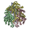
- Structure visualization
| Structure viewer | Molecule: MolmilJmol/JSmol |
|---|
- Downloads & links
-Download
| PDBx/mmCIF format | 1f8i.cif.gz | 353 KB | Display | PDBx/mmCIF format |
|---|---|---|---|---|
| PDB format | pdb1f8i.ent.gz | 286.5 KB | Display | PDB format |
| PDBx/mmJSON format | 1f8i.json.gz | Tree view | PDBx/mmJSON format | |
| Others |  Other downloads Other downloads |
-Validation report
| Arichive directory | https://data.pdbj.org/pub/pdb/validation_reports/f8/1f8iftp://data.pdbj.org/pub/pdb/validation_reports/f8/1f8i | HTTPS FTP |
|---|
-Related structure data


-Links
 PDBj
PDBj
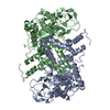
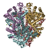
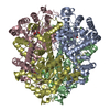
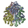
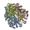
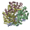
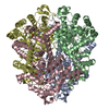
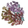
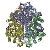
- Assembly
| Deposited unit | 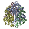
| ||||||||
|---|---|---|---|---|---|---|---|---|---|
| 1 |
| ||||||||
| Unit cell |
|
-Components
| #1: Protein | Mass: 47189.410 Da / Num. of mol.: 4 / Mutation: C191S Source method: isolated from a genetically manipulated source Source: (gene. exp.) Mycobacterium tuberculosis H37Rv (bacteria)Species: Mycobacterium tuberculosis / Strain: H37RV / Plasmid: PET30B / Production host: References: UniProt: P0A5H3, UniProt: P9WKK7*PLUS, isocitrate lyase #2: Chemical | ChemComp-MG /   Mass: 24.305 Da / Num. of mol.: 4 / Source method: obtained synthetically / Formula: Mg Mass: 24.305 Da / Num. of mol.: 4 / Source method: obtained synthetically / Formula: Mg#3: Chemical | ChemComp-GLV / 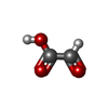  Mass: 74.035 Da / Num. of mol.: 4 / Source method: obtained synthetically / Formula: C2H2O3 Mass: 74.035 Da / Num. of mol.: 4 / Source method: obtained synthetically / Formula: C2H2O3#4: Chemical | ChemComp-SIN /   Mass: 118.088 Da / Num. of mol.: 4 / Source method: obtained synthetically / Formula: C4H6O4 Mass: 118.088 Da / Num. of mol.: 4 / Source method: obtained synthetically / Formula: C4H6O4#5: Water | ChemComp-HOH / |  Mass: 18.015 Da / Num. of mol.: 1052 / Source method: isolated from a natural source / Formula: H2O Mass: 18.015 Da / Num. of mol.: 1052 / Source method: isolated from a natural source / Formula: H2O |
|---|
-Experimental details
-Experiment
| Experiment | Method: X-RAY DIFFRACTION / Number of used crystals: 1 |
|---|
- Sample preparation
| Crystal | Density Matthews: 2.12 Å3/Da / Density % sol: 41.95 % | ||||||||||||||||||||
|---|---|---|---|---|---|---|---|---|---|---|---|---|---|---|---|---|---|---|---|---|---|
| Crystal grow | Temperature: 289 K / Method: microbatch under paraffin oil / pH: 8 Details: PEG4000, sodium acetate, Tris.HCl, magnesium acetate, glyoxylate, 3-nitropropionate, pH 8.0, Microbatch under Paraffin oil, temperature 289.0K | ||||||||||||||||||||
| Crystal grow | *PLUS Method: vapor diffusion | ||||||||||||||||||||
| Components of the solutions | *PLUS
|
-Data collection
| Diffraction | Mean temperature: 100 K |
|---|---|
| Diffraction source | Source: SYNCHROTRON / Site: APS  / Beamline: 14-BM-C / Wavelength: 1 / Beamline: 14-BM-C / Wavelength: 1 |
| Detector | Type: ADSC QUANTUM 4 / Detector: CCD / Date: Mar 3, 2000 |
| Radiation | Protocol: SINGLE WAVELENGTH / Monochromatic (M) / Laue (L): M / Scattering type: x-ray |
| Radiation wavelength | Wavelength: 1 Å / Relative weight: 1 |
| Reflection | Resolution: 2.25→99 Å / Num. all: 75213 / Num. obs: 72007 / % possible obs: 92.8 % / Observed criterion σ(F): 0 / Observed criterion σ(I): 1 / Redundancy: 4.12 % / Biso Wilson estimate: 19.8 Å2 / Rmerge(I) obs: 0.05 / Net I/σ(I): 18.6 |
| Reflection shell | Resolution: 2.25→2.33 Å / Redundancy: 3 % / Rmerge(I) obs: 0.122 / Num. unique all: 6128 / % possible all: 79.8 |
| Reflection | *PLUS Num. measured all: 296878 / Rmerge(I) obs: 0.05 |
| Reflection shell | *PLUS % possible obs: 79.8 % |
- Processing
| Software |
| ||||||||||||||||||||||||||||||||||||||||
|---|---|---|---|---|---|---|---|---|---|---|---|---|---|---|---|---|---|---|---|---|---|---|---|---|---|---|---|---|---|---|---|---|---|---|---|---|---|---|---|---|---|
| Refinement | Resolution: 2.25→35.02 Å / Rfactor Rfree error: 0.002 / Data cutoff high absF: 199873.12 / Data cutoff low absF: 0 / Isotropic thermal model: RESTRAINED / Cross valid method: THROUGHOUT / σ(F): 0 / σ(I): 0 / Stereochemistry target values: Engh & Huber
| ||||||||||||||||||||||||||||||||||||||||
| Solvent computation | Solvent model: FLAT MODEL / Bsol: 42.37 Å2 / ksol: 0.358 e/Å3 | ||||||||||||||||||||||||||||||||||||||||
| Displacement parameters | Biso mean: 26.6 Å2
| ||||||||||||||||||||||||||||||||||||||||
| Refine analyze |
| ||||||||||||||||||||||||||||||||||||||||
| Refinement step | Cycle: LAST / Resolution: 2.25→35.02 Å
| ||||||||||||||||||||||||||||||||||||||||
| Refine LS restraints |
| ||||||||||||||||||||||||||||||||||||||||
| Refine LS restraints NCS | NCS model details: CONSTR | ||||||||||||||||||||||||||||||||||||||||
| LS refinement shell | Resolution: 2.25→2.39 Å / Rfactor Rfree error: 0.008 / Total num. of bins used: 6
| ||||||||||||||||||||||||||||||||||||||||
| Xplor file |
| ||||||||||||||||||||||||||||||||||||||||
| Software | *PLUS Name: CNS / Version: 1 / Classification: refinement | ||||||||||||||||||||||||||||||||||||||||
| Refinement | *PLUS σ(F): 0 / % reflection Rfree: 10.1 % / Rfactor Rfree: 0.21 | ||||||||||||||||||||||||||||||||||||||||
| Solvent computation | *PLUS | ||||||||||||||||||||||||||||||||||||||||
| Displacement parameters | *PLUS Biso mean: 26.6 Å2 | ||||||||||||||||||||||||||||||||||||||||
| Refine LS restraints | *PLUS
| ||||||||||||||||||||||||||||||||||||||||
| LS refinement shell | *PLUS Rfactor Rfree: 0.249 / % reflection Rfree: 10 % / Rfactor Rwork: 0.192 |
