 Movie
Movie Controller


+ Open data

- Basic information
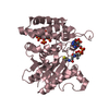
| Entry | Database: PDB / ID: 1f5m | ||||||
|---|---|---|---|---|---|---|---|
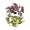
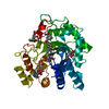
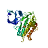
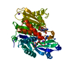
| Title | STRUCTURE OF THE GAF DOMAIN | ||||||
 Components Components | GAF | ||||||
 Keywords Keywords | SIGNALING PROTEIN / GAF / cGMP binding | ||||||
| Function / homology |  Function and homology information Function and homology informationL-methionine (R)-S-oxide reductase / L-methionine (R)-S-oxide reductase activity / cellular response to oxidative stress / nucleus / cytoplasm / cytosol Similarity search - Function | ||||||
| Biological species |  | ||||||
| Method |  X-RAY DIFFRACTION / SYNCHROTRON / Resolution: 1.9 Å X-RAY DIFFRACTION / SYNCHROTRON / Resolution: 1.9 Å | ||||||
 Authors Authors | Ho, Y.S. / Burden, L.M. / Hurley, J.H. | ||||||
 Citation Citation | Journal: EMBO J. / Year: 2000 Title: Structure of the GAF domain, a ubiquitous signaling motif and a new class of cyclic GMP receptor. Authors: Ho, Y.S. / Burden, L.M. / Hurley, J.H. | ||||||
| History |
|
- Structure visualization
| Structure viewer | Molecule: MolmilJmol/JSmol |
|---|
- Downloads & links
-Download
| PDBx/mmCIF format | 1f5m.cif.gz | 86.4 KB | Display | PDBx/mmCIF format |
|---|---|---|---|---|
| PDB format | pdb1f5m.ent.gz | 65.8 KB | Display | PDB format |
| PDBx/mmJSON format | 1f5m.json.gz | Tree view | PDBx/mmJSON format | |
| Others |  Other downloads Other downloads |
-Validation report
| Arichive directory | https://data.pdbj.org/pub/pdb/validation_reports/f5/1f5mftp://data.pdbj.org/pub/pdb/validation_reports/f5/1f5m | HTTPS FTP |
|---|
-Related structure data
| Similar structure data |
|---|


-Links
 PDBj
PDBj


- Assembly
| Deposited unit | 
| ||||||||
|---|---|---|---|---|---|---|---|---|---|
| 1 |
| ||||||||
| 2 | 
| ||||||||
| 3 | 
| ||||||||
| Unit cell |
|
-Components
| #1: Protein | Mass: 19753.252 Da / Num. of mol.: 2 Source method: isolated from a genetically manipulated source Source: (gene. exp.) Production host:  #2: Chemical | ChemComp-BR /   Mass: 79.904 Da / Num. of mol.: 9 / Source method: obtained synthetically / Formula: Br Mass: 79.904 Da / Num. of mol.: 9 / Source method: obtained synthetically / Formula: Br#3: Water | ChemComp-HOH / |  Mass: 18.015 Da / Num. of mol.: 346 / Source method: isolated from a natural source / Formula: H2O Mass: 18.015 Da / Num. of mol.: 346 / Source method: isolated from a natural source / Formula: H2OHas protein modification | Y | |
|---|
-Experimental details
-Experiment
| Experiment | Method: X-RAY DIFFRACTION / Number of used crystals: 1 |
|---|
- Sample preparation
| Crystal | Density Matthews: 2.79 Å3/Da / Density % sol: 55.87 % | ||||||||||||||||||||||||||||||||||||||||||
|---|---|---|---|---|---|---|---|---|---|---|---|---|---|---|---|---|---|---|---|---|---|---|---|---|---|---|---|---|---|---|---|---|---|---|---|---|---|---|---|---|---|---|---|
| Crystal grow | Temperature: 295 K / Method: vapor diffusion, hanging drop / pH: 8 Details: ammonium sulfate, lithium sulfate, Tris buffer, pH 8, VAPOR DIFFUSION, HANGING DROP, temperature 295K | ||||||||||||||||||||||||||||||||||||||||||
| Crystal grow | *PLUS pH: 8 | ||||||||||||||||||||||||||||||||||||||||||
| Components of the solutions | *PLUS
|
-Data collection
| Diffraction | Mean temperature: 100 K |
|---|---|
| Diffraction source | Source: SYNCHROTRON / Site: NSLS  / Beamline: X9B / Wavelength: 0.915 / Beamline: X9B / Wavelength: 0.915 |
| Detector | Type: ADSC QUANTUM 4 / Detector: CCD / Date: Jan 23, 2000 |
| Radiation | Protocol: SINGLE WAVELENGTH / Monochromatic (M) / Laue (L): M / Scattering type: x-ray |
| Radiation wavelength | Wavelength: 0.915 Å / Relative weight: 1 |
| Reflection | Resolution: 1.9→20 Å / Num. all: 67128 / Num. obs: 36203 / % possible obs: 99.7 % / Observed criterion σ(F): 2 / Observed criterion σ(I): 2 / Redundancy: 1.8 % / Biso Wilson estimate: 25.3 Å2 / Rmerge(I) obs: 0.057 / Net I/σ(I): 27.9 |
| Reflection shell | Resolution: 1.9→1.97 Å / Rmerge(I) obs: 0.545 / % possible all: 99.5 |
| Reflection | *PLUS Num. measured all: 901143 |
| Reflection shell | *PLUS Highest resolution: 1.9 Å / % possible obs: 99.5 % / Mean I/σ(I) obs: 2.73 |
- Processing
| Software |
| ||||||||||||||||||||||||||||
|---|---|---|---|---|---|---|---|---|---|---|---|---|---|---|---|---|---|---|---|---|---|---|---|---|---|---|---|---|---|
| Refinement | Resolution: 1.9→500 Å
| ||||||||||||||||||||||||||||
| Displacement parameters |
| ||||||||||||||||||||||||||||
| Refinement step | Cycle: LAST / Resolution: 1.9→500 Å
| ||||||||||||||||||||||||||||
| Refine LS restraints |
| ||||||||||||||||||||||||||||
| Xplor file |
| ||||||||||||||||||||||||||||
| Software | *PLUS Name: CNS / Classification: refinement | ||||||||||||||||||||||||||||
| Refinement | *PLUS Highest resolution: 1.9 Å / Lowest resolution: 500 Å | ||||||||||||||||||||||||||||
| Solvent computation | *PLUS | ||||||||||||||||||||||||||||
| Displacement parameters | *PLUS | ||||||||||||||||||||||||||||
| Refine LS restraints | *PLUS
|
