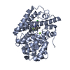
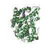
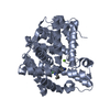
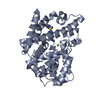
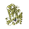
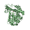
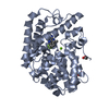
Entry Database : PDB / ID : 6b97Title Crystal structure of PDE2 in complex with complex 9 cGMP-dependent 3',5'-cyclic phosphodiesterase Keywords / Function / homology Function Domain/homology Component
/ / / / / / / / / / / / / / / / / / / / / / / / / / / / / / / / / / / / / / / / / / / / / / / / / / / / / / / / / / / / / / / / / / / / / / / / / / / / / / / / / / / / Biological species Homo sapiens (human)Method / / Resolution : 1.76 Å Authors Lu, J. Journal : Bioorg. Med. Chem. Lett. / Year : 2017Title : The identification of a novel lead class for phosphodiesterase 2 inhibition by fragment-based drug design.Authors: Forster, A.B. / Abeywickrema, P. / Bunda, J. / Cox, C.D. / Cabalu, T.D. / Egbertson, M. / Fay, J. / Getty, K. / Hall, D. / Kornienko, M. / Lu, J. / Parthasarathy, G. / Reid, J. / Sharma, S. ... Authors : Forster, A.B. / Abeywickrema, P. / Bunda, J. / Cox, C.D. / Cabalu, T.D. / Egbertson, M. / Fay, J. / Getty, K. / Hall, D. / Kornienko, M. / Lu, J. / Parthasarathy, G. / Reid, J. / Sharma, S. / Shipe, W.D. / Smith, S.M. / Soisson, S. / Stachel, S.J. / Su, H.P. / Wang, D. / Berger, R. History Deposition Oct 10, 2017 Deposition site / Processing site Revision 1.0 Nov 22, 2017 Provider / Type Revision 1.1 Nov 29, 2017 Group / Category Item / _citation.page_first / _citation.page_lastRevision 1.2 Mar 6, 2024 Group Data collection / Database references ... Data collection / Database references / Derived calculations / Refinement description Category chem_comp_atom / chem_comp_bond ... chem_comp_atom / chem_comp_bond / database_2 / pdbx_struct_conn_angle / software / struct_conn Item _database_2.pdbx_DOI / _database_2.pdbx_database_accession ... _database_2.pdbx_DOI / _database_2.pdbx_database_accession / _pdbx_struct_conn_angle.ptnr1_auth_seq_id / _pdbx_struct_conn_angle.ptnr3_auth_seq_id / _pdbx_struct_conn_angle.value / _software.name / _struct_conn.pdbx_dist_value / _struct_conn.ptnr1_auth_asym_id / _struct_conn.ptnr1_auth_comp_id / _struct_conn.ptnr1_auth_seq_id / _struct_conn.ptnr1_label_asym_id / _struct_conn.ptnr1_label_atom_id / _struct_conn.ptnr1_label_comp_id / _struct_conn.ptnr1_label_seq_id / _struct_conn.ptnr2_auth_asym_id / _struct_conn.ptnr2_auth_comp_id / _struct_conn.ptnr2_auth_seq_id / _struct_conn.ptnr2_label_asym_id / _struct_conn.ptnr2_label_atom_id / _struct_conn.ptnr2_label_comp_id
Show all Show less
 Movie
Movie Controller


 Open data
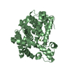
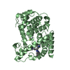
 Basic information
 Components
 Keywords
 Function and homology information
 Homo sapiens (human)
 X-RAY DIFFRACTION /
  Authors
 Citation
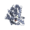
 Structure visualization
 Downloads & links
 Other downloads


 PDBj
PDBj

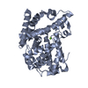
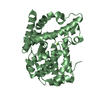
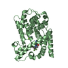
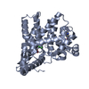
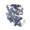
 Assembly
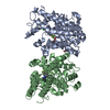


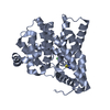
 Spodoptera frugiperda (fall armyworm)


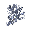
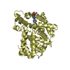
 Mass: 65.409 Da / Num. of mol.: 2 / Source method: obtained synthetically / Formula: Zn
 Mass: 24.305 Da / Num. of mol.: 2 / Source method: obtained synthetically / Formula: Mg
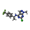
 Mass: 355.745 Da / Num. of mol.: 2 / Source method: obtained synthetically / Formula: C15H13ClF3N5
 Mass: 62.068 Da / Num. of mol.: 2 / Source method: obtained synthetically / Formula: C2H6O2
 Sample preparation
 / Beamline: 17-ID / Wavelength: 1 Å
 Processing