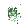
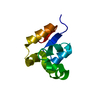
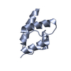
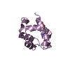
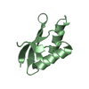
Entry Database : PDB / ID : 1f0mTitle MONOMERIC STRUCTURE OF THE HUMAN EPHB2 SAM (STERILE ALPHA MOTIF) DOMAIN EPHRIN TYPE-B RECEPTOR 2 Keywords / Function / homology Function Domain/homology Component
/ / / / / / / / / / / / / / / / / / / / / / / / / / / / / / / / / / / / / / / / / / / / / / / / / / / / / / / / / / / / / / / / / / / / / / / / / / / / / / / / / / / / / / / / / / / / / / / / / / / / / / / / / / / / / / / / / / / / / / / / / / / / / / / / / / / / / / / / / / / / / / / Biological species Homo sapiens (human)Method / / Resolution : 2.2 Å Authors Thanos, C.D. / Faham, S. / Goodwill, K.E. / Cascio, D. / Phillips, M. / Bowie, J.U. Journal : J.Biol.Chem. / Year : 1999Title : Monomeric structure of the human EphB2 sterile alpha motif domain.Authors : Thanos, C.D. / Faham, S. / Goodwill, K.E. / Cascio, D. / Phillips, M. / Bowie, J.U. History Deposition May 16, 2000 Deposition site / Processing site Revision 1.0 Jul 4, 2000 Provider / Type Revision 1.1 Apr 27, 2008 Group Revision 1.2 Jul 13, 2011 Group Revision 1.3 Jan 31, 2018 Group / Category / Item Revision 1.4 Feb 7, 2024 Group / Database referencesCategory chem_comp_atom / chem_comp_bond ... chem_comp_atom / chem_comp_bond / database_2 / struct_ref_seq_dif Item / _database_2.pdbx_database_accession / _struct_ref_seq_dif.details
Show all Show less
 Movie
Movie Controller

 Yorodumi
 Open data
 Basic information
 Components
 Keywords
 Function and homology information
 Homo sapiens (human)
 X-RAY DIFFRACTION /
  Authors
 Citation
 Structure visualization
 Downloads & links
 Other downloads
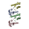


 PDBj
PDBj


 Assembly
 Mass: 18.015 Da / Num. of mol.: 113 / Source method: isolated from a natural source / Formula: H2O
 Sample preparation
 / Beamline: X12C / Wavelength: 1
  Processing