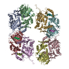
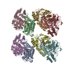
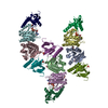
Entry Database : PDB / ID : 1b4fTitle OLIGOMERIC STRUCTURE OF THE HUMAN EPHB2 RECEPTOR SAM DOMAIN EPHB2 Keywords / / / Function / homology Function Domain/homology Component
/ / / / / / / / / / / / / / / / / / / / / / / / / / / / / / / / / / / / / / / / / / / / / / / / / / / / / / / / / / / / / / / / / / / / / / / / / / / / / / / / / / / / / / / / / / / / / / / / / / / / / / / / / / / / / / / / / / / / / / / / / / / / / / / / / / / / / / / / / / / / / / / Biological species Homo sapiens (human)Method / / / Resolution : 1.95 Å Authors Thanos, C.D. / Goodwill, K.E. / Bowie, J.U. Journal : Science / Year : 1999Title : Oligomeric structure of the human EphB2 receptor SAM domain.Authors : Thanos, C.D. / Goodwill, K.E. / Bowie, J.U. History Deposition Dec 20, 1998 Processing site Revision 1.0 Feb 16, 1999 Provider / Type Revision 1.1 Mar 24, 2008 Group Revision 1.2 Jul 13, 2011 Group Revision 1.3 Dec 11, 2019 Group / Database references / Derived calculationsCategory pdbx_distant_solvent_atoms / pdbx_struct_assembly ... pdbx_distant_solvent_atoms / pdbx_struct_assembly / pdbx_struct_assembly_gen / pdbx_struct_oper_list / struct_ref_seq_dif Item Revision 1.4 Feb 7, 2024 Group / Database references / Category / chem_comp_bond / database_2Item / _database_2.pdbx_database_accession
Show all Show less
 Movie
Movie Controller


 Open data
 Basic information
 Components
 Keywords
 Function and homology information
 Homo sapiens (human)
 X-RAY DIFFRACTION /
  Authors
 Citation
 Structure visualization
 Downloads & links
 Other downloads
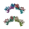

 PDBj
PDBj


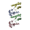
 Assembly


 Mass: 18.015 Da / Num. of mol.: 685 / Source method: isolated from a natural source / Formula: H2O
 Sample preparation
 / Beamline: 5.0.2 / Wavelength: 0.96859, 0.97966, 0.97982, 1.0000
 Processing