 Movie
Movie Controller


+ Open data

- Basic information
| Entry | Database: PDB / ID: 1eth | |||||||||
|---|---|---|---|---|---|---|---|---|---|---|
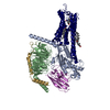
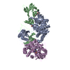
| Title | TRIACYLGLYCEROL LIPASE/COLIPASE COMPLEX | |||||||||
 Components Components |
| |||||||||
 Keywords Keywords | COMPLEX (HYDROLASE/COFACTOR) / COMPLEX (HYDROLASE-COFACTOR) / LIPID DEGRADATION / COMPLEX (HYDROLASE-COFACTOR) complex | |||||||||
| Function / homology |  Function and homology information Function and homology informationDigestion of dietary lipid / Retinoid metabolism and transport / all-trans-retinyl-palmitate hydrolase, all-trans-retinol forming activity / lipoprotein lipase activity / lipase binding / glycerophospholipid phospholipase A1 activity / triglyceride catabolic process / triacylglycerol lipase / high-density lipoprotein particle remodeling / triacylglycerol lipase activity ...Digestion of dietary lipid / Retinoid metabolism and transport / all-trans-retinyl-palmitate hydrolase, all-trans-retinol forming activity / lipoprotein lipase activity / lipase binding / glycerophospholipid phospholipase A1 activity / triglyceride catabolic process / triacylglycerol lipase / high-density lipoprotein particle remodeling / triacylglycerol lipase activity / response to food / retinoid metabolic process / digestion / lipid catabolic process / cholesterol homeostasis / response to bacterium / enzyme activator activity / fatty acid biosynthetic process / : / extracellular region / metal ion binding Similarity search - Function | |||||||||
| Biological species |  | |||||||||
| Method |  X-RAY DIFFRACTION / SYNCHROTRON / Resolution: 2.8 Å X-RAY DIFFRACTION / SYNCHROTRON / Resolution: 2.8 Å | |||||||||
 Authors Authors | Hermoso, J. / Pignol, D. / Kerfelec, B. / Crenon, I. / Chapus, C. / Fontecilla-Camps, J.C. | |||||||||
 Citation Citation | Journal: J.Biol.Chem. / Year: 1996 Title: Lipase activation by nonionic detergents. The crystal structure of the porcine lipase-colipase-tetraethylene glycol monooctyl ether complex. Authors: Hermoso, J. / Pignol, D. / Kerfelec, B. / Crenon, I. / Chapus, C. / Fontecilla-Camps, J.C. #1: Journal: Nature / Year: 1993Title: Interfacial Activation of the Lipase-Procolipase Complex by Mixed Micelles Revealed by X-Ray Crystallography Authors: Van Tilbeurgh, H. / Egloff, M.P. / Martinez, C. / Rugani, N. / Verger, R. / Cambillau, C. #2: Journal: Nature / Year: 1992Title: Structure of the Pancreatic Lipase-Procolipase Complex Authors: Van Tilbeurgh, H. / Sarda, L. / Verger, R. / Cambillau, C. | |||||||||
| History |
|
- Structure visualization
| Structure viewer | Molecule: MolmilJmol/JSmol |
|---|
- Downloads & links
-Download
| PDBx/mmCIF format | 1eth.cif.gz | 288.3 KB | Display | PDBx/mmCIF format |
|---|---|---|---|---|
| PDB format | pdb1eth.ent.gz | 235.7 KB | Display | PDB format |
| PDBx/mmJSON format | 1eth.json.gz | Tree view | PDBx/mmJSON format | |
| Others |  Other downloads Other downloads |
-Validation report
| Arichive directory | https://data.pdbj.org/pub/pdb/validation_reports/et/1ethftp://data.pdbj.org/pub/pdb/validation_reports/et/1eth | HTTPS FTP |
|---|
-Related structure data
| Similar structure data |
|---|


-Links
 PDBj
PDBj


- Assembly
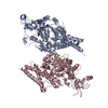
| Deposited unit | 
| ||||||||
|---|---|---|---|---|---|---|---|---|---|
| 1 | 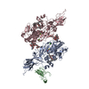
| ||||||||
| 2 | 
| ||||||||
| Unit cell |
|
-Components
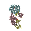
-Protein , 2 types, 4 molecules ACBD
| #1: Protein | Mass: 49908.863 Da / Num. of mol.: 2 / Source method: isolated from a natural source / Source: (natural) #2: Protein | Mass: 10319.651 Da / Num. of mol.: 2 / Source method: isolated from a natural source / Source: (natural) |
|---|
-Sugars , 1 types, 2 molecules
| #3: Polysaccharide | Source method: isolated from a genetically manipulated source |
|---|
-Non-polymers , 4 types, 365 molecules 


| #4: Chemical |  Mass: 40.078 Da / Num. of mol.: 2 / Source method: obtained synthetically / Formula: Ca Mass: 40.078 Da / Num. of mol.: 2 / Source method: obtained synthetically / Formula: Ca#5: Chemical | ChemComp-C8E / (  Mass: 306.438 Da / Num. of mol.: 4 / Source method: obtained synthetically / Formula: C16H34O5 / Comment: C8E, detergent*YM Mass: 306.438 Da / Num. of mol.: 4 / Source method: obtained synthetically / Formula: C16H34O5 / Comment: C8E, detergent*YM#6: Chemical |  Mass: 78.133 Da / Num. of mol.: 2 / Source method: obtained synthetically / Formula: C2H6OS Mass: 78.133 Da / Num. of mol.: 2 / Source method: obtained synthetically / Formula: C2H6OS#7: Water | ChemComp-HOH / | Mass: 18.015 Da / Num. of mol.: 357 / Source method: isolated from a natural source / Formula: H2O |
|---|
-Details
| Has protein modification | Y |
|---|
-Experimental details
-Experiment
| Experiment | Method: X-RAY DIFFRACTION |
|---|
- Sample preparation
| Crystal | Density Matthews: 4.18 Å3/Da / Density % sol: 61 % | ||||||||||||||||||||||||||||||||||||
|---|---|---|---|---|---|---|---|---|---|---|---|---|---|---|---|---|---|---|---|---|---|---|---|---|---|---|---|---|---|---|---|---|---|---|---|---|---|
| Crystal | *PLUS Density % sol: 70 % | ||||||||||||||||||||||||||||||||||||
| Crystal grow | *PLUS Temperature: 20 ℃ / pH: 6 / Method: vapor diffusion, sitting drop | ||||||||||||||||||||||||||||||||||||
| Components of the solutions | *PLUS
|
-Data collection
| Diffraction source | Source: SYNCHROTRON / Site: LURE  / Beamline: DW32 / Wavelength: 0.9 / Beamline: DW32 / Wavelength: 0.9 |
|---|---|
| Detector | Detector: IMAGE PLATE |
| Radiation | Monochromatic (M) / Laue (L): M / Scattering type: x-ray |
| Radiation wavelength | Wavelength: 0.9 Å / Relative weight: 1 |
| Reflection | Resolution: 2.8→12.4 Å / Num. obs: 42501 / % possible obs: 86 % / Observed criterion σ(I): 0 / Redundancy: 4 % / Rmerge(I) obs: 0.13 |
- Processing
| Software |
| ||||||||||||||||||||||||||||||||||||||||||||||||||||||||||||
|---|---|---|---|---|---|---|---|---|---|---|---|---|---|---|---|---|---|---|---|---|---|---|---|---|---|---|---|---|---|---|---|---|---|---|---|---|---|---|---|---|---|---|---|---|---|---|---|---|---|---|---|---|---|---|---|---|---|---|---|---|---|
| Refinement | Resolution: 2.8→6 Å / σ(F): 2 / Details: CRYST1 UNUSUAL UNIT-CELL DATA: PSEUDOSYMMETRY P 23
| ||||||||||||||||||||||||||||||||||||||||||||||||||||||||||||
| Displacement parameters | Biso mean: 19.73 Å2 | ||||||||||||||||||||||||||||||||||||||||||||||||||||||||||||
| Refinement step | Cycle: LAST / Resolution: 2.8→6 Å
| ||||||||||||||||||||||||||||||||||||||||||||||||||||||||||||
| Refine LS restraints |
| ||||||||||||||||||||||||||||||||||||||||||||||||||||||||||||
| Software | *PLUS Name: X-PLOR / Classification: refinement | ||||||||||||||||||||||||||||||||||||||||||||||||||||||||||||
| Refinement | *PLUS | ||||||||||||||||||||||||||||||||||||||||||||||||||||||||||||
| Solvent computation | *PLUS | ||||||||||||||||||||||||||||||||||||||||||||||||||||||||||||
| Displacement parameters | *PLUS | ||||||||||||||||||||||||||||||||||||||||||||||||||||||||||||
| Refine LS restraints | *PLUS
|
