 Movie
Movie Controller

[English] 日本語
 Yorodumi
- PDB-1ecr: ESCHERICHIA COLI REPLICATION TERMINATOR PROTEIN (TUS) COMPLEXED W... -
+ Open data

- Basic information
| Entry | Database: PDB / ID: 1ecr | ||||||
|---|---|---|---|---|---|---|---|
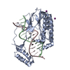
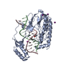
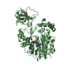
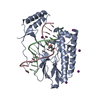
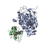
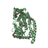
| Title | ESCHERICHIA COLI REPLICATION TERMINATOR PROTEIN (TUS) COMPLEXED WITH DNA | ||||||
 Components Components |
| ||||||
 Keywords Keywords | REPLICATION/DNA / DNA-BINDING / DNA REPLICATION / COMPLEX (DNA-BINDING PROTEIN-DNA) / REPLICATION-DNA COMPLEX | ||||||
| Function / homology |  Function and homology information Function and homology informationreplication fork arrest involved in DNA replication termination / DNA replication termination / sequence-specific DNA binding / cytoplasm Similarity search - Function | ||||||
| Biological species |  | ||||||
| Method |  X-RAY DIFFRACTION / SYNCHROTRON / MIR, ANOMALOUS SCATTERING / Resolution: 2.7 Å X-RAY DIFFRACTION / SYNCHROTRON / MIR, ANOMALOUS SCATTERING / Resolution: 2.7 Å | ||||||
 Authors Authors | Kamada, K. / Morikawa, K. | ||||||
 Citation Citation | Journal: Nature / Year: 1996 Title: Structure of a replication-terminator protein complexed with DNA. Authors: Kamada, K. / Horiuchi, T. / Ohsumi, K. / Shimamoto, N. / Morikawa, K. #1: Journal: Proteins / Year: 1996Title: Crystallization and Preliminary X-Ray Analysis of the Escherichia Coli Replication Terminator Protein Complexed with DNA Authors: Kamada, K. / Ohsumi, K. / Horiuchi, T. / Shimamoto, N. / Morikawa, K. | ||||||
| History |
|
- Structure visualization
| Structure viewer | Molecule: MolmilJmol/JSmol |
|---|
- Downloads & links
-Download
| PDBx/mmCIF format | 1ecr.cif.gz | 93.9 KB | Display | PDBx/mmCIF format |
|---|---|---|---|---|
| PDB format | pdb1ecr.ent.gz | 68.9 KB | Display | PDB format |
| PDBx/mmJSON format | 1ecr.json.gz | Tree view | PDBx/mmJSON format | |
| Others |  Other downloads Other downloads |
-Validation report
| Arichive directory | https://data.pdbj.org/pub/pdb/validation_reports/ec/1ecrftp://data.pdbj.org/pub/pdb/validation_reports/ec/1ecr | HTTPS FTP |
|---|
-Related structure data
| Similar structure data |
|---|


-Links
 PDBj
PDBj


- Assembly
| Deposited unit | 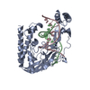
| ||||||||
|---|---|---|---|---|---|---|---|---|---|
| 1 |
| ||||||||
| Unit cell |
|
-Components
| #1: DNA chain | Mass: 4856.191 Da / Num. of mol.: 1 / Source method: obtained synthetically |
|---|---|
| #2: DNA chain | Mass: 4927.225 Da / Num. of mol.: 1 / Source method: obtained synthetically |
| #3: Protein | Mass: 35843.188 Da / Num. of mol.: 1 Source method: isolated from a genetically manipulated source Source: (gene. exp.) |
| #4: Water | ChemComp-HOH /  Mass: 18.015 Da / Num. of mol.: 49 / Source method: isolated from a natural source / Formula: H2O Mass: 18.015 Da / Num. of mol.: 49 / Source method: isolated from a natural source / Formula: H2O |
-Experimental details
-Experiment
| Experiment | Method: X-RAY DIFFRACTION |
|---|
- Sample preparation
| Crystal | Density Matthews: 2.9 Å3/Da / Density % sol: 57 % | |||||||||||||||||||||||||||||||||||||||||||||||||||||||
|---|---|---|---|---|---|---|---|---|---|---|---|---|---|---|---|---|---|---|---|---|---|---|---|---|---|---|---|---|---|---|---|---|---|---|---|---|---|---|---|---|---|---|---|---|---|---|---|---|---|---|---|---|---|---|---|---|
| Crystal grow | Temperature: 293 K / Method: microdialysis / pH: 5.3 / Details: pH 5.30, MICRODIALYSIS, temperature 293.00K | |||||||||||||||||||||||||||||||||||||||||||||||||||||||
| Components of the solutions |
| |||||||||||||||||||||||||||||||||||||||||||||||||||||||
| Crystal | *PLUS | |||||||||||||||||||||||||||||||||||||||||||||||||||||||
| Crystal grow | *PLUS pH: 5.3 Details: Kamada, K., (1996) Proteins: Struct.,Funct., Genet., 24, 402. | |||||||||||||||||||||||||||||||||||||||||||||||||||||||
| Components of the solutions | *PLUS
|
-Data collection
| Diffraction | Mean temperature: 288 K |
|---|---|
| Diffraction source | Source: SYNCHROTRON / Site: Photon Factory  / Beamline: BL-6A / Beamline: BL-6A |
| Detector | Type: FUJI / Detector: IMAGE PLATE / Date: Dec 13, 1994 / Details: TOROIDAL MIRROR |
| Radiation | Monochromator: SI(111) / Protocol: SINGLE WAVELENGTH / Monochromatic (M) / Laue (L): M / Scattering type: x-ray |
| Radiation wavelength | Relative weight: 1 |
| Reflection | Resolution: 2.7→50 Å / Num. obs: 14018 / % possible obs: 90.4 % / Observed criterion σ(F): 1 / Observed criterion σ(I): 1 / Redundancy: 3.3 % / Rsym value: 0.06 |
| Reflection shell | Resolution: 2.7→2.8 Å / Redundancy: 2.6 % / Rsym value: 0.217 / % possible all: 75.1 |
| Reflection | *PLUS Highest resolution: 2.7 Å / Lowest resolution: 50 Å / % possible obs: 90.4 % / Rmerge(I) obs: 0.06 |
- Processing
| Software |
| ||||||||||||
|---|---|---|---|---|---|---|---|---|---|---|---|---|---|
| Refinement | Method to determine structure: MIR, ANOMALOUS SCATTERING / Resolution: 2.7→10 Å / σ(F): 0
| ||||||||||||
| Refinement step | Cycle: LAST / Resolution: 2.7→10 Å
| ||||||||||||
| Refinement | *PLUS Highest resolution: 2.7 Å / Lowest resolution: 10 Å / σ(F): 0 | ||||||||||||
| Solvent computation | *PLUS | ||||||||||||
| Displacement parameters | *PLUS |
