 Movie
Movie Controller

[English] 日本語
 Yorodumi
- PDB-2i05: Escherichia Coli Replication Terminator Protein (Tus) Complexed W... -
+ Open data

- Basic information
| Entry | Database: PDB / ID: 2i05 | ||||||
|---|---|---|---|---|---|---|---|
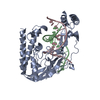
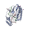
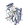
| Title | Escherichia Coli Replication Terminator Protein (Tus) Complexed With TerA DNA | ||||||
 Components Components |
| ||||||
 Keywords Keywords | REPLICATION/DNA / PROTEIN-DNA COMPLEX / REPLICATION-DNA COMPLEX | ||||||
| Function / homology |  Function and homology information Function and homology informationreplication fork arrest involved in DNA replication termination / DNA replication termination / sequence-specific DNA binding / cytoplasm Similarity search - Function | ||||||
| Biological species |  | ||||||
| Method |  X-RAY DIFFRACTION / SYNCHROTRON / MOLECULAR REPLACEMENT / Resolution: 2.6 Å X-RAY DIFFRACTION / SYNCHROTRON / MOLECULAR REPLACEMENT / Resolution: 2.6 Å | ||||||
 Authors Authors | Oakley, A.J. / Mulcair, M.D. / Schaeffer, P.M. / Dixon, N.E. | ||||||
 Citation Citation | Journal: To be Published Title: Polarity of Termination of DNA Replication in E. coli Authors: Oakley, A.J. / Mulcair, M.D. / Schaeffer, P.M. / Dixon, N.E. #1: Journal: CELL(CAMBRIDGE,MASS.) / Year: 2006Title: A molecular mousetrap determines polarity of termination of DNA replication in E. coli Authors: Mulcair, M.D. / Schaeffer, P.M. / Oakley, A.J. / Cross, H.F. / Neylon, C. / Hill, T.M. / Dixon, N.E. | ||||||
| History |
|
- Structure visualization
| Structure viewer | Molecule: MolmilJmol/JSmol |
|---|
- Downloads & links
-Download
| PDBx/mmCIF format | 2i05.cif.gz | 94.9 KB | Display | PDBx/mmCIF format |
|---|---|---|---|---|
| PDB format | pdb2i05.ent.gz | 68.7 KB | Display | PDB format |
| PDBx/mmJSON format | 2i05.json.gz | Tree view | PDBx/mmJSON format | |
| Others |  Other downloads Other downloads |
-Validation report
| Arichive directory | https://data.pdbj.org/pub/pdb/validation_reports/i0/2i05ftp://data.pdbj.org/pub/pdb/validation_reports/i0/2i05 | HTTPS FTP |
|---|
-Related structure data
| Related structure data | 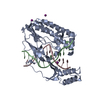 2ewjS S: Starting model for refinement |
|---|---|
| Similar structure data |


-Links
 PDBj
PDBj


- Assembly
| Deposited unit | 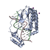
| ||||||||
|---|---|---|---|---|---|---|---|---|---|
| 1 |
| ||||||||
| Unit cell |
|
-Components
| #1: DNA chain | Mass: 4856.191 Da / Num. of mol.: 1 / Source method: obtained synthetically | ||
|---|---|---|---|
| #2: DNA chain | Mass: 4927.225 Da / Num. of mol.: 1 / Source method: obtained synthetically | ||
| #3: Protein | Mass: 35843.188 Da / Num. of mol.: 1 / Fragment: TUS Source method: isolated from a genetically manipulated source Source: (gene. exp.) | ||
| #4: Chemical |   Mass: 126.904 Da / Num. of mol.: 2 / Source method: obtained synthetically / Formula: I Mass: 126.904 Da / Num. of mol.: 2 / Source method: obtained synthetically / Formula: I#5: Water | ChemComp-HOH / |  Mass: 18.015 Da / Num. of mol.: 10 / Source method: isolated from a natural source / Formula: H2O Mass: 18.015 Da / Num. of mol.: 10 / Source method: isolated from a natural source / Formula: H2O |
-Experimental details
-Experiment
| Experiment | Method: X-RAY DIFFRACTION / Number of used crystals: 1 |
|---|
- Sample preparation
| Crystal | Density Matthews: 2.76 Å3/Da / Density % sol: 55.44 % | ||||||||||||||||||||||||||||
|---|---|---|---|---|---|---|---|---|---|---|---|---|---|---|---|---|---|---|---|---|---|---|---|---|---|---|---|---|---|
| Crystal grow | Temperature: 298 K / Method: vapor diffusion, hanging drop / pH: 6.5 Details: 12.5% PEG 3350, 50mM Bis-Tris pH6.5, 200mM NaI, VAPOR DIFFUSION, HANGING DROP, temperature 298K | ||||||||||||||||||||||||||||
| Components of the solutions |
|
-Data collection
| Diffraction | Mean temperature: 100 K |
|---|---|
| Diffraction source | Source: SYNCHROTRON / Site: APS  / Beamline: 19-ID / Wavelength: 0.97929 Å / Beamline: 19-ID / Wavelength: 0.97929 Å |
| Detector | Type: ADSC QUANTUM 315 / Detector: CCD / Date: Mar 2, 2006 Details: Rosenbaum-Rock monochromator, high-resolution double-crystal sagittal focusing, double crystal, Rosenbaum-Rock vertical focusing mirror |
| Radiation | Monochromator: Rosenbaum-Rock / Protocol: SINGLE WAVELENGTH / Monochromatic (M) / Laue (L): M / Scattering type: x-ray |
| Radiation wavelength | Wavelength: 0.97929 Å / Relative weight: 1 |
| Reflection | Resolution: 2.6→50 Å / Num. obs: 13969 / % possible obs: 82 % / Observed criterion σ(F): -2 / Observed criterion σ(I): -2 / Redundancy: 5.2 % / Biso Wilson estimate: 56.028 Å2 / Rmerge(I) obs: 0.088 / Rsym value: 0.088 / Net I/σ(I): 28.36 |
| Reflection shell | Resolution: 2.6→2.69 Å / Redundancy: 2.2 % / Rmerge(I) obs: 0.259 / Mean I/σ(I) obs: 3.1 / Num. unique all: 421 / Rsym value: 0.259 / % possible all: 25.5 |
- Processing
| Software |
| ||||||||||||||||||||||||||||||||||||||||||||||||||||||||||||||||||||||||||||||||||||||||||||||||||||||||||||||||||||||||||||||||||||||||||||||||||||||||||||||||||||||||||
|---|---|---|---|---|---|---|---|---|---|---|---|---|---|---|---|---|---|---|---|---|---|---|---|---|---|---|---|---|---|---|---|---|---|---|---|---|---|---|---|---|---|---|---|---|---|---|---|---|---|---|---|---|---|---|---|---|---|---|---|---|---|---|---|---|---|---|---|---|---|---|---|---|---|---|---|---|---|---|---|---|---|---|---|---|---|---|---|---|---|---|---|---|---|---|---|---|---|---|---|---|---|---|---|---|---|---|---|---|---|---|---|---|---|---|---|---|---|---|---|---|---|---|---|---|---|---|---|---|---|---|---|---|---|---|---|---|---|---|---|---|---|---|---|---|---|---|---|---|---|---|---|---|---|---|---|---|---|---|---|---|---|---|---|---|---|---|---|---|---|---|---|
| Refinement | Method to determine structure: MOLECULAR REPLACEMENT Starting model: 2EWJ Resolution: 2.6→50 Å / Cor.coef. Fo:Fc: 0.932 / Cor.coef. Fo:Fc free: 0.87 / SU B: 29.777 / SU ML: 0.287 / TLS residual ADP flag: LIKELY RESIDUAL / Cross valid method: THROUGHOUT / σ(F): -2 / ESU R: 1.345 / ESU R Free: 0.429 / Stereochemistry target values: MAXIMUM LIKELIHOOD
| ||||||||||||||||||||||||||||||||||||||||||||||||||||||||||||||||||||||||||||||||||||||||||||||||||||||||||||||||||||||||||||||||||||||||||||||||||||||||||||||||||||||||||
| Solvent computation | Ion probe radii: 0.8 Å / Shrinkage radii: 0.8 Å / VDW probe radii: 1.2 Å / Solvent model: BABINET MODEL WITH MASK | ||||||||||||||||||||||||||||||||||||||||||||||||||||||||||||||||||||||||||||||||||||||||||||||||||||||||||||||||||||||||||||||||||||||||||||||||||||||||||||||||||||||||||
| Displacement parameters | Biso mean: 53.288 Å2
| ||||||||||||||||||||||||||||||||||||||||||||||||||||||||||||||||||||||||||||||||||||||||||||||||||||||||||||||||||||||||||||||||||||||||||||||||||||||||||||||||||||||||||
| Refinement step | Cycle: LAST / Resolution: 2.6→50 Å
| ||||||||||||||||||||||||||||||||||||||||||||||||||||||||||||||||||||||||||||||||||||||||||||||||||||||||||||||||||||||||||||||||||||||||||||||||||||||||||||||||||||||||||
| Refine LS restraints |
| ||||||||||||||||||||||||||||||||||||||||||||||||||||||||||||||||||||||||||||||||||||||||||||||||||||||||||||||||||||||||||||||||||||||||||||||||||||||||||||||||||||||||||
| LS refinement shell | Resolution: 2.6→2.668 Å / Total num. of bins used: 20
| ||||||||||||||||||||||||||||||||||||||||||||||||||||||||||||||||||||||||||||||||||||||||||||||||||||||||||||||||||||||||||||||||||||||||||||||||||||||||||||||||||||||||||
| Refinement TLS params. | Method: refined / Origin x: 38.127 Å / Origin y: 4.4711 Å / Origin z: 50.221 Å
|
