 Movie
Movie Controller


+ Open data

- Basic information
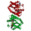
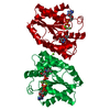
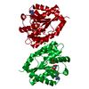
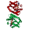
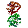
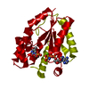
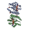
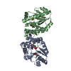
| Entry | Database: PDB / ID: 1e9f | ||||||
|---|---|---|---|---|---|---|---|
| Title | Mutant human thymidylate kinase complexed with TMP and ADP | ||||||
 Components Components | THYMIDYLATE KINASE | ||||||
 Keywords Keywords | TRANSFERASE / PHOSPHOTRANSFERASE / THYMIDYLATE KINASE / P-LOOP | ||||||
| Function / homology |  Function and homology information Function and homology informationthymidine biosynthetic process / dTMP kinase / dUDP biosynthetic process / dTDP biosynthetic process / dTMP kinase activity / Interconversion of nucleotide di- and triphosphates / nucleoside diphosphate kinase activity / dTTP biosynthetic process / cellular response to growth factor stimulus / mitochondrion ...thymidine biosynthetic process / dTMP kinase / dUDP biosynthetic process / dTDP biosynthetic process / dTMP kinase activity / Interconversion of nucleotide di- and triphosphates / nucleoside diphosphate kinase activity / dTTP biosynthetic process / cellular response to growth factor stimulus / mitochondrion / ATP binding / nucleus / cytoplasm / cytosol Similarity search - Function | ||||||
| Biological species |  HOMO SAPIENS (human) HOMO SAPIENS (human) | ||||||
| Method |  X-RAY DIFFRACTION / SYNCHROTRON / MOLECULAR REPLACEMENT / Resolution: 1.9 Å X-RAY DIFFRACTION / SYNCHROTRON / MOLECULAR REPLACEMENT / Resolution: 1.9 Å | ||||||
 Authors Authors | Ostermann, N. / Lavie, A. / Padiyar, S. / Brundiers, R. / Veit, T. / Reinstein, J. / Goody, R.S. / Konrad, M. / Schlichting, I. | ||||||
 Citation Citation | Journal: J.Mol.Biol. / Year: 2000 Title: Potentiating Azt Activation: Structures of Wildtype and Mutant Human Thymidylate Kinase Suggest Reasons for the Mutants' Improved Kinetics with the HIV Prodrug Metabolite Aztmp Authors: Ostermann, N. / Lavie, A. / Padiyar, S. / Brundiers, R. / Veit, T. / Reintein, J. / Goody, R.S. / Konrad, M. / Schlichting, I. #1: Journal: Structure / Year: 2000Title: Insights Into the Phosphoryltransfer Mechanism of Human Thymidylate Kinase Gained from Crystal Structures of Enzyme Complexes Along the Reaction Coordinate. Authors: Ostermann, N. / Schlichting, I. / Brundiers, R. / Konrad, M. / Reinstein, J. / Veit, T. / Goody, R.S. / Lavie, A. | ||||||
| History |
|
- Structure visualization
| Structure viewer | Molecule: MolmilJmol/JSmol |
|---|
- Downloads & links
-Download
| PDBx/mmCIF format | 1e9f.cif.gz | 61.2 KB | Display | PDBx/mmCIF format |
|---|---|---|---|---|
| PDB format | pdb1e9f.ent.gz | 43.9 KB | Display | PDB format |
| PDBx/mmJSON format | 1e9f.json.gz | Tree view | PDBx/mmJSON format | |
| Others |  Other downloads Other downloads |
-Validation report
| Arichive directory | https://data.pdbj.org/pub/pdb/validation_reports/e9/1e9fftp://data.pdbj.org/pub/pdb/validation_reports/e9/1e9f | HTTPS FTP |
|---|
-Related structure data
| Related structure data | 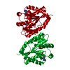 1e98C 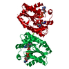 1e99C  1e9aC 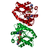 1e9bC 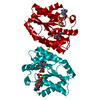 1e9cC 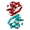 1e9dC 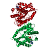 1e9eC C: citing same article ( |
|---|---|
| Similar structure data |


-Links
 PDBj
PDBj

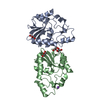
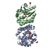
- Assembly
| Deposited unit | 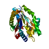
| ||||||||
|---|---|---|---|---|---|---|---|---|---|
| 1 | 
| ||||||||
| Unit cell |
| ||||||||
| Components on special symmetry positions |
|
-Components
| #1: Protein | Mass: 24223.729 Da / Num. of mol.: 1 / Mutation: YES Source method: isolated from a genetically manipulated source Source: (gene. exp.) HOMO SAPIENS (human), (gene. exp.) Production host: | ||||||
|---|---|---|---|---|---|---|---|
| #2: Chemical | ChemComp-TMP / 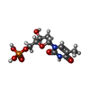  Mass: 322.208 Da / Num. of mol.: 1 / Source method: obtained synthetically / Formula: C10H15N2O8P Mass: 322.208 Da / Num. of mol.: 1 / Source method: obtained synthetically / Formula: C10H15N2O8P | ||||||
| #3: Chemical | ChemComp-ADP /   Mass: 427.201 Da / Num. of mol.: 1 / Source method: obtained synthetically / Formula: C10H15N5O10P2 / Comment: ADP, energy-carrying molecule*YM Mass: 427.201 Da / Num. of mol.: 1 / Source method: obtained synthetically / Formula: C10H15N5O10P2 / Comment: ADP, energy-carrying molecule*YM | ||||||
| #4: Chemical |   Mass: 24.305 Da / Num. of mol.: 2 / Source method: obtained synthetically / Formula: Mg Mass: 24.305 Da / Num. of mol.: 2 / Source method: obtained synthetically / Formula: Mg#5: Water | ChemComp-HOH / |  Mass: 18.015 Da / Num. of mol.: 162 / Source method: isolated from a natural source / Formula: H2O Mass: 18.015 Da / Num. of mol.: 162 / Source method: isolated from a natural source / Formula: H2OCompound details | CHAIN A - ENGINEERED MUTATION ARG200ALA, ARG16GLY CHAIN A - (GLY SER HIS) INSERTED AT THE N- ...CHAIN A - ENGINEERED | Sequence details | SAMPLE COMTAINS SER183, ILE184, ASP190, AND A ILE191 (CONFIRMED BY THE DNA SEQUENCE AND ELECTRON ...SAMPLE COMTAINS SER183, ILE184, ASP190, AND A ILE191 (CONFIRMED BY THE DNA SEQUENCE AND ELECTRON DENSITY OF SIDE-CHAINS). POSSIBLE ERROR IN SWISSPROT ENTRY. | |
-Experimental details
-Experiment
| Experiment | Method: X-RAY DIFFRACTION / Number of used crystals: 1 |
|---|
- Sample preparation
| Crystal | Density Matthews: 2.55 Å3/Da / Density % sol: 51.75 % | ||||||||||||||||||||||||||||||||||||||||||||||||||||||
|---|---|---|---|---|---|---|---|---|---|---|---|---|---|---|---|---|---|---|---|---|---|---|---|---|---|---|---|---|---|---|---|---|---|---|---|---|---|---|---|---|---|---|---|---|---|---|---|---|---|---|---|---|---|---|---|
| Crystal grow | Temperature: 293 K / Method: vapor diffusion / pH: 8 Details: VAPOR DIFFUSION AT 293 K MIXING 2 MICROLITERS OF A SOLUTION CONTAINING THE ENZYME, NUCLEOTIDES, AND 50 MM MGCL2 WITH 2 MICROLITERS OF A SOLUTION CONTAINING 21% PEG 3350, 100 MM TRIS HCL PH 8. ...Details: VAPOR DIFFUSION AT 293 K MIXING 2 MICROLITERS OF A SOLUTION CONTAINING THE ENZYME, NUCLEOTIDES, AND 50 MM MGCL2 WITH 2 MICROLITERS OF A SOLUTION CONTAINING 21% PEG 3350, 100 MM TRIS HCL PH 8.0 AND 50 MICROLITERS OF DEAD SEA WATER. | ||||||||||||||||||||||||||||||||||||||||||||||||||||||
| Crystal grow | *PLUS Temperature: 20 ℃ / Method: vapor diffusion, hanging drop / Details: Ostermann, N., (2000) Structure (London), 8, 629. | ||||||||||||||||||||||||||||||||||||||||||||||||||||||
| Components of the solutions | *PLUS
|
-Data collection
| Diffraction | Mean temperature: 100 K |
|---|---|
| Diffraction source | Source: SYNCHROTRON / Site: EMBL/DESY, HAMBURG  / Beamline: BW7B / Wavelength: 0.8443 / Beamline: BW7B / Wavelength: 0.8443 |
| Radiation | Monochromator: YES / Protocol: SINGLE WAVELENGTH / Monochromatic (M) / Laue (L): M / Scattering type: x-ray |
| Radiation wavelength | Wavelength: 0.8443 Å / Relative weight: 1 |
| Reflection | Resolution: 1.9→70 Å / Num. obs: 19646 / % possible obs: 96.8 % / Redundancy: 4.9 % / Rsym value: 0.054 / Net I/σ(I): 17 |
| Reflection shell | Resolution: 1.9→2 Å / Redundancy: 4.3 % / Mean I/σ(I) obs: 3.2 / Rsym value: 0.144 / % possible all: 97.3 |
| Reflection | *PLUS Lowest resolution: 70 Å / Num. measured all: 96858 / Rmerge(I) obs: 0.054 |
| Reflection shell | *PLUS % possible obs: 97.3 % / Rmerge(I) obs: 0.144 |
- Processing
| Software |
| ||||||||||||||||||||
|---|---|---|---|---|---|---|---|---|---|---|---|---|---|---|---|---|---|---|---|---|---|
| Refinement | Method to determine structure: MOLECULAR REPLACEMENT / Resolution: 1.9→70 Å / Cross valid method: THROUGHOUT / σ(F): 0
| ||||||||||||||||||||
| Refinement step | Cycle: LAST / Resolution: 1.9→70 Å
| ||||||||||||||||||||
| Software | *PLUS Name: REFMAC / Classification: refinement | ||||||||||||||||||||
| Refinement | *PLUS Lowest resolution: 70 Å / Rfactor obs: 0.226 | ||||||||||||||||||||
| Solvent computation | *PLUS | ||||||||||||||||||||
| Displacement parameters | *PLUS | ||||||||||||||||||||
| Refine LS restraints | *PLUS
|
