Mass: 18.015 Da / Num. of mol.: 297 / Source method: isolated from a natural source / Formula: H2O
-
Details
Compound details
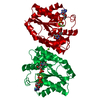
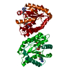
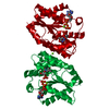
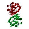
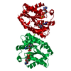
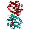
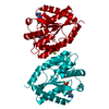
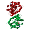
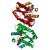
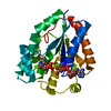
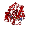
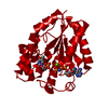
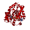
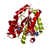
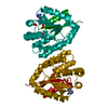
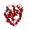
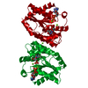
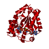
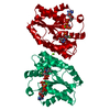
CHAIN A ENGINEERED MUTATION ARG200ALA GLY, SER, HIS INSERTED AT THE N-TERMINUS THE ACTIVE SITE ...CHAIN A ENGINEERED MUTATION ARG200ALA GLY, SER, HIS INSERTED AT THE N-TERMINUS THE ACTIVE SITE CONTAINS A MIXTURE OF NUCLEOTIDES: TMP ADP AND TMP + ANP, THE ORIENTATION OF TMP DIFFERS.
Sequence details
SAMPLE COMTAINS SER183, ILE184, ASP190, AND A ILE191 (CONFIRMED BY THE DNA SEQUENCE AND ELECTRON ...SAMPLE COMTAINS SER183, ILE184, ASP190, AND A ILE191 (CONFIRMED BY THE DNA SEQUENCE AND ELECTRON DENSITY OF SIDE-CHAINS). POSSIBLE ERROR IN SWISSPROT ENTRY.
-
Experimental details
-
Experiment
Experiment
Method: X-RAY DIFFRACTION / Number of used crystals: 1
-
Sample preparation
Crystal
Density Matthews: 2.63 Å3/Da / Density % sol: 53.22 %
Crystal grow
Temperature: 293 K / Method: vapor diffusion / pH: 8 Details: VAPOR DIFFUSION AT 293 K MIXING 2 MICROLITERS OF A SOLUTION CONTAINING THE ENZYME, NUCLEOTIDES, AND 50 MM MGCL2 WITH 2 MICROLITERS OF A SOLUTION CONTAINING 21% PEG 3350, 100 MM TRIS HCL PH 8. ...Details: VAPOR DIFFUSION AT 293 K MIXING 2 MICROLITERS OF A SOLUTION CONTAINING THE ENZYME, NUCLEOTIDES, AND 50 MM MGCL2 WITH 2 MICROLITERS OF A SOLUTION CONTAINING 21% PEG 3350, 100 MM TRIS HCL PH 8.0 AND 50 MICROLITERS OF DEAD SEA WATER.
Crystal grow
*PLUS
Temperature: 20 ℃ / Method: vapor diffusion, hanging drop
In the structure databanks used in Yorodumi, some data are registered as the other names, "COVID-19 virus" and "2019-nCoV". Here are the details of the virus and the list of structure data.
Jan 31, 2019. EMDB accession codes are about to change! (news from PDBe EMDB page)
EMDB accession codes are about to change! (news from PDBe EMDB page)
The allocation of 4 digits for EMDB accession codes will soon come to an end. Whilst these codes will remain in use, new EMDB accession codes will include an additional digit and will expand incrementally as the available range of codes is exhausted. The current 4-digit format prefixed with “EMD-” (i.e. EMD-XXXX) will advance to a 5-digit format (i.e. EMD-XXXXX), and so on. It is currently estimated that the 4-digit codes will be depleted around Spring 2019, at which point the 5-digit format will come into force.
The EM Navigator/Yorodumi systems omit the EMD- prefix.
Related info.:Q: What is EMD? / ID/Accession-code notation in Yorodumi/EM Navigator
Yorodumi is a browser for structure data from EMDB, PDB, SASBDB, etc.
This page is also the successor to EM Navigator detail page, and also detail information page/front-end page for Omokage search.
The word "yorodu" (or yorozu) is an old Japanese word meaning "ten thousand". "mi" (miru) is to see.
Related info.:EMDB / PDB / SASBDB / Comparison of 3 databanks / Yorodumi Search / Aug 31, 2016. New EM Navigator & Yorodumi / Yorodumi Papers / Jmol/JSmol / Function and homology information / Changes in new EM Navigator and Yorodumi
 Movie
Movie Controller

 Yorodumi
 Open data
 Basic information
 Components
 Keywords
 Function and homology information
 HOMO SAPIENS (human)
 X-RAY DIFFRACTION /
  Authors
 Citation
 Structure visualization
 Downloads & links
 Other downloads


 PDBj
PDBj


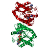
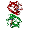
 Assembly


 Mass: 322.208 Da / Num. of mol.: 1 / Source method: obtained synthetically / Formula: C10H15N2O8P
 Mass: 506.196 Da / Num. of mol.: 1 / Source method: obtained synthetically / Formula: C10H17N6O12P3 / Comment: AMP-PNP, energy-carrying molecule analogue*YM
 Mass: 427.201 Da / Num. of mol.: 1 / Source method: obtained synthetically / Formula: C10H15N5O10P2 / Comment: ADP, energy-carrying molecule*YM
 Mass: 24.305 Da / Num. of mol.: 2 / Source method: obtained synthetically / Formula: Mg
 Sample preparation
 / Beamline: BW7B / Wavelength: 0.847
  Processing