 Movie
Movie Controller


+ Open data

- Basic information
| Entry | Database: PDB / ID: 1nn3 | ||||||
|---|---|---|---|---|---|---|---|
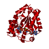
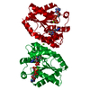
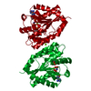
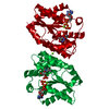
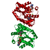
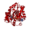
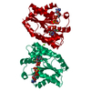
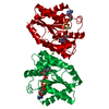
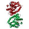
| Title | Crystal structure of human thymidylate kinase with d4TMP + ADP | ||||||
 Components Components | similar to THYMIDYLATE KINASE (DTMP KINASE) | ||||||
 Keywords Keywords | TRANSFERASE / thymidylate kinase / p-loop / d4TMP | ||||||
| Function / homology |  Function and homology information Function and homology informationthymidine biosynthetic process / dTMP kinase / dUDP biosynthetic process / dTDP biosynthetic process / dTMP kinase activity / Interconversion of nucleotide di- and triphosphates / nucleoside diphosphate kinase activity / dTTP biosynthetic process / cellular response to growth factor stimulus / mitochondrion ...thymidine biosynthetic process / dTMP kinase / dUDP biosynthetic process / dTDP biosynthetic process / dTMP kinase activity / Interconversion of nucleotide di- and triphosphates / nucleoside diphosphate kinase activity / dTTP biosynthetic process / cellular response to growth factor stimulus / mitochondrion / ATP binding / nucleus / cytoplasm / cytosol Similarity search - Function | ||||||
| Biological species |  Homo sapiens (human) Homo sapiens (human) | ||||||
| Method |  X-RAY DIFFRACTION / SYNCHROTRON / FOURIER SYNTHESIS / Resolution: 1.55 Å X-RAY DIFFRACTION / SYNCHROTRON / FOURIER SYNTHESIS / Resolution: 1.55 Å | ||||||
 Authors Authors | Ostermann, N. / Segura-Pena, D. / Meier, C. / Veit, T. / Monnerjahn, M. / Konrad, M. / Lavie, A. | ||||||
 Citation Citation | Journal: Biochemistry / Year: 2003 Title: Structures of human thymidylate kinase in complex with prodrugs: implications for the structure-based design of novel compounds Authors: Ostermann, N. / Segura-Pena, D. / Meier, C. / Veit, T. / Monnerjahn, M. / Konrad, M. / Lavie, A. | ||||||
| History |
|
- Structure visualization
| Structure viewer | Molecule: MolmilJmol/JSmol |
|---|
- Downloads & links
-Download
| PDBx/mmCIF format | 1nn3.cif.gz | 68.1 KB | Display | PDBx/mmCIF format |
|---|---|---|---|---|
| PDB format | pdb1nn3.ent.gz | 49.1 KB | Display | PDB format |
| PDBx/mmJSON format | 1nn3.json.gz | Tree view | PDBx/mmJSON format | |
| Others |  Other downloads Other downloads |
-Validation report
| Arichive directory | https://data.pdbj.org/pub/pdb/validation_reports/nn/1nn3ftp://data.pdbj.org/pub/pdb/validation_reports/nn/1nn3 | HTTPS FTP |
|---|
-Related structure data
| Related structure data | 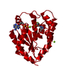 1nmxC 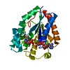 1nmyC  1nmzC  1nn0C 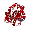 1nn1C 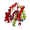 1nn5C C: citing same article ( |
|---|---|
| Similar structure data |

-Links
 PDBj
PDBj

- Assembly
| Deposited unit | 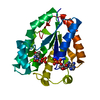
| ||||||||
|---|---|---|---|---|---|---|---|---|---|
| 1 | 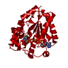
| ||||||||
| Unit cell |
|
-Components
| #1: Protein | Mass: 24052.533 Da / Num. of mol.: 1 / Mutation: R200A Source method: isolated from a genetically manipulated source Source: (gene. exp.) Homo sapiens (human) / Production host:  | ||||||
|---|---|---|---|---|---|---|---|
| #2: Chemical |   Mass: 24.305 Da / Num. of mol.: 2 / Source method: obtained synthetically / Formula: Mg Mass: 24.305 Da / Num. of mol.: 2 / Source method: obtained synthetically / Formula: Mg#3: Chemical | ChemComp-2DT / |   Type: DNA linking / Mass: 306.209 Da / Num. of mol.: 1 / Source method: obtained synthetically / Formula: C10H15N2O7P Type: DNA linking / Mass: 306.209 Da / Num. of mol.: 1 / Source method: obtained synthetically / Formula: C10H15N2O7P#4: Chemical | ChemComp-ADP / |   Mass: 427.201 Da / Num. of mol.: 1 / Source method: obtained synthetically / Formula: C10H15N5O10P2 / Comment: ADP, energy-carrying molecule*YM Mass: 427.201 Da / Num. of mol.: 1 / Source method: obtained synthetically / Formula: C10H15N5O10P2 / Comment: ADP, energy-carrying molecule*YM#5: Water | ChemComp-HOH / |  Mass: 18.015 Da / Num. of mol.: 315 / Source method: isolated from a natural source / Formula: H2O Mass: 18.015 Da / Num. of mol.: 315 / Source method: isolated from a natural source / Formula: H2O |
-Experimental details
-Experiment
| Experiment | Method: X-RAY DIFFRACTION / Number of used crystals: 1 |
|---|
- Sample preparation
| Crystal | Density Matthews: 2.12 Å3/Da / Density % sol: 41.65 % | |||||||||||||||
|---|---|---|---|---|---|---|---|---|---|---|---|---|---|---|---|---|
| Crystal grow | Temperature: 293 K / Method: vapor diffusion, hanging drop / pH: 8 Details: 15-20% PEG 3350, 100 mM Tris/HCl, pH 8.0, 5% filtered dead sea water, VAPOR DIFFUSION, HANGING DROP, temperature 293K | |||||||||||||||
| Crystal grow | *PLUS | |||||||||||||||
| Components of the solutions | *PLUS
|
-Data collection
| Diffraction | Mean temperature: 100 K |
|---|---|
| Diffraction source | Source: SYNCHROTRON / Site: EMBL/DESY, HAMBURG  / Beamline: X11 / Wavelength: 0.9076 Å / Beamline: X11 / Wavelength: 0.9076 Å |
| Detector | Type: MARRESEARCH / Detector: IMAGE PLATE |
| Radiation | Protocol: SINGLE WAVELENGTH / Monochromatic (M) / Laue (L): M / Scattering type: x-ray |
| Radiation wavelength | Wavelength: 0.9076 Å / Relative weight: 1 |
| Reflection | Resolution: 1.55→35.7 Å / Num. obs: 37349 / % possible obs: 98.4 % / Redundancy: 4.4 % / Rmerge(I) obs: 0.042 / Net I/σ(I): 40.3 |
| Reflection shell | Resolution: 1.55→1.6 Å / Redundancy: 4 % / Mean I/σ(I) obs: 8.5 / Rsym value: 0.322 / % possible all: 96.8 |
| Reflection | *PLUS Num. measured all: 165795 |
| Reflection shell | *PLUS % possible obs: 96.8 % / Rmerge(I) obs: 0.322 |
- Processing
| Software |
| |||||||||||||||
|---|---|---|---|---|---|---|---|---|---|---|---|---|---|---|---|---|
| Refinement | Method to determine structure: FOURIER SYNTHESIS / Resolution: 1.55→35.7 Å / σ(F): 0
| |||||||||||||||
| Refinement step | Cycle: LAST / Resolution: 1.55→35.7 Å
| |||||||||||||||
| Refinement | *PLUS Num. reflection obs: 37350 / % reflection Rfree: 10 % | |||||||||||||||
| Solvent computation | *PLUS | |||||||||||||||
| Displacement parameters | *PLUS | |||||||||||||||
| Refine LS restraints | *PLUS
|
