 Movie
Movie Controller


+ Open data

- Basic information
| Entry | Database: PDB / ID: 1.0E+87 | ||||||
|---|---|---|---|---|---|---|---|
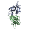
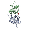
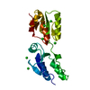
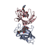
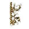
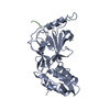
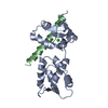
| Title | Human CD69 - trigonal form | ||||||
 Components Components | EARLY ACTIVATION ANTIGEN CD69 | ||||||
 Keywords Keywords | SUGAR BINDING PROTEIN / HEMATOPOIETIC CELL RECEPTOR / LEUCOCYTE / NKD / KLR | ||||||
| Function / homology |  Function and homology information Function and homology informationnegative regulation of T cell migration / negative regulation of T-helper 17 cell lineage commitment / molecular sequestering activity / negative regulation of inflammatory response / cellular response to xenobiotic stimulus / transmembrane signaling receptor activity / carbohydrate binding / external side of plasma membrane / protein-containing complex / identical protein binding / plasma membrane Similarity search - Function | ||||||
| Biological species |  HOMO SAPIENS (human) HOMO SAPIENS (human) | ||||||
| Method |  X-RAY DIFFRACTION / SYNCHROTRON / MOLECULAR REPLACEMENT / Resolution: 1.5 Å X-RAY DIFFRACTION / SYNCHROTRON / MOLECULAR REPLACEMENT / Resolution: 1.5 Å | ||||||
 Authors Authors | Tormo, J. | ||||||
 Citation Citation | Journal: J.Biol.Chem. / Year: 2001 Title: Crystal Structure of the C-Type Lectin-Like Domain from the Human Hematopoietic Cell Receptor Cd69 Authors: Llera, A.S. / Viedma, F. / Sanchez-Madrid, F. / Tormo, J. | ||||||
| History |
|
- Structure visualization
| Structure viewer | Molecule: MolmilJmol/JSmol |
|---|
- Downloads & links
-Download
| PDBx/mmCIF format | 1e87.cif.gz | 41.9 KB | Display | PDBx/mmCIF format |
|---|---|---|---|---|
| PDB format | pdb1e87.ent.gz | 28.2 KB | Display | PDB format |
| PDBx/mmJSON format | 1e87.json.gz | Tree view | PDBx/mmJSON format | |
| Others |  Other downloads Other downloads |
-Validation report
| Arichive directory | https://data.pdbj.org/pub/pdb/validation_reports/e8/1e87ftp://data.pdbj.org/pub/pdb/validation_reports/e8/1e87 | HTTPS FTP |
|---|
-Related structure data
| Related structure data |  1e8iSC S: Starting model for refinement C: citing same article ( |
|---|---|
| Similar structure data |

-Links
 PDBj
PDBj


- Assembly
| Deposited unit | 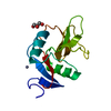
| ||||||||
|---|---|---|---|---|---|---|---|---|---|
| 1 | 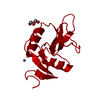
| ||||||||
| Unit cell |
| ||||||||
| Details | THE BIOLOGICALLY FUNTIONAL MOLECULE IS A DIMER WHICH CAN BE BUILT BY APPLYING THE MATRIX GIVEN IN REMARK 350 |
-Components
| #1: Protein | Mass: 13806.557 Da / Num. of mol.: 1 / Fragment: C-TYPE LECTIN-LIKE DOMAIN, RESIDUES 82-199 Source method: isolated from a genetically manipulated source Source: (gene. exp.) HOMO SAPIENS (human) / Cell: HEMATOPOIETIC CELL / Cellular location: CELL SURFACE / Cellular location (production host): INCLUSION BODIES / Production host:  |
|---|---|
| #2: Chemical | ChemComp-ZN /   Mass: 65.409 Da / Num. of mol.: 1 / Source method: obtained synthetically / Formula: Zn Mass: 65.409 Da / Num. of mol.: 1 / Source method: obtained synthetically / Formula: Zn |
| #3: Chemical | ChemComp-GOL /   Mass: 92.094 Da / Num. of mol.: 1 / Source method: obtained synthetically / Formula: C3H8O3 Mass: 92.094 Da / Num. of mol.: 1 / Source method: obtained synthetically / Formula: C3H8O3 |
| #4: Water | ChemComp-HOH /  Mass: 18.015 Da / Num. of mol.: 108 / Source method: isolated from a natural source / Formula: H2O Mass: 18.015 Da / Num. of mol.: 108 / Source method: isolated from a natural source / Formula: H2O |
| Compound details | SIGNAL TRANSMITTING RECEPTOR IN LYMPHOCYTES, NATURAL KILLER (NK) CELLS, AND PLATELETS. INVOLVED IN ...SIGNAL TRANSMITTI |
| Has protein modification | Y |
-Experimental details
-Experiment
| Experiment | Method: X-RAY DIFFRACTION / Number of used crystals: 1 |
|---|
- Sample preparation
| Crystal | Density Matthews: 2.94 Å3/Da / Density % sol: 58 % | |||||||||||||||||||||||||||||||||||||||||||||||||
|---|---|---|---|---|---|---|---|---|---|---|---|---|---|---|---|---|---|---|---|---|---|---|---|---|---|---|---|---|---|---|---|---|---|---|---|---|---|---|---|---|---|---|---|---|---|---|---|---|---|---|
| Crystal grow | pH: 5.2 / Details: pH 5.20 | |||||||||||||||||||||||||||||||||||||||||||||||||
| Crystal grow | *PLUS pH: 7.2 / Method: vapor diffusion | |||||||||||||||||||||||||||||||||||||||||||||||||
| Components of the solutions | *PLUS
|
-Data collection
| Diffraction | Mean temperature: 100 K |
|---|---|
| Diffraction source | Source: SYNCHROTRON / Site: ESRF  / Beamline: ID14-3 / Wavelength: 0.931 / Beamline: ID14-3 / Wavelength: 0.931 |
| Detector | Type: MARRESEARCH / Detector: CCD |
| Radiation | Protocol: SINGLE WAVELENGTH / Monochromatic (M) / Laue (L): M / Scattering type: x-ray |
| Radiation wavelength | Wavelength: 0.931 Å / Relative weight: 1 |
| Reflection | Resolution: 1.5→20 Å / Num. obs: 26542 / % possible obs: 98.6 % / Redundancy: 5.7 % / Biso Wilson estimate: 19.5 Å2 / Rsym value: 0.062 / Net I/σ(I): 33.5 |
| Reflection shell | Resolution: 1.5→1.53 Å / Mean I/σ(I) obs: 15.6 / Rsym value: 0.106 / % possible all: 100 |
| Reflection | *PLUS Num. measured all: 151066 / Rmerge(I) obs: 0.062 |
| Reflection shell | *PLUS % possible obs: 100 % / Rmerge(I) obs: 0.106 |
- Processing
| Software |
| ||||||||||||||||||||||||||||||||||||||||||||||||||||||||||||||||||||||||||||||||
|---|---|---|---|---|---|---|---|---|---|---|---|---|---|---|---|---|---|---|---|---|---|---|---|---|---|---|---|---|---|---|---|---|---|---|---|---|---|---|---|---|---|---|---|---|---|---|---|---|---|---|---|---|---|---|---|---|---|---|---|---|---|---|---|---|---|---|---|---|---|---|---|---|---|---|---|---|---|---|---|---|---|
| Refinement | Method to determine structure: MOLECULAR REPLACEMENT Starting model: PDB ENTRY 1E8I Resolution: 1.5→19.79 Å / Rfactor Rfree error: 0.006 / Isotropic thermal model: RESTRAINED / Cross valid method: THROUGHOUT / σ(F): 0
| ||||||||||||||||||||||||||||||||||||||||||||||||||||||||||||||||||||||||||||||||
| Solvent computation | Solvent model: FLAT MODEL / Bsol: 35.6827 Å2 / ksol: 0.386021 e/Å3 | ||||||||||||||||||||||||||||||||||||||||||||||||||||||||||||||||||||||||||||||||
| Displacement parameters | Biso mean: 20.3 Å2
| ||||||||||||||||||||||||||||||||||||||||||||||||||||||||||||||||||||||||||||||||
| Refine analyze |
| ||||||||||||||||||||||||||||||||||||||||||||||||||||||||||||||||||||||||||||||||
| Refinement step | Cycle: LAST / Resolution: 1.5→19.79 Å
| ||||||||||||||||||||||||||||||||||||||||||||||||||||||||||||||||||||||||||||||||
| Refine LS restraints |
| ||||||||||||||||||||||||||||||||||||||||||||||||||||||||||||||||||||||||||||||||
| LS refinement shell | Resolution: 1.5→1.59 Å / Rfactor Rfree error: 0.015 / Total num. of bins used: 6
| ||||||||||||||||||||||||||||||||||||||||||||||||||||||||||||||||||||||||||||||||
| Xplor file |
| ||||||||||||||||||||||||||||||||||||||||||||||||||||||||||||||||||||||||||||||||
| Software | *PLUS Name: CNS / Version: 1 / Classification: refinement | ||||||||||||||||||||||||||||||||||||||||||||||||||||||||||||||||||||||||||||||||
| Refinement | *PLUS Lowest resolution: 20 Å / Num. reflection obs: 24561 / % reflection Rfree: 7.3 % / Rfactor Rfree: 0.244 | ||||||||||||||||||||||||||||||||||||||||||||||||||||||||||||||||||||||||||||||||
| Solvent computation | *PLUS | ||||||||||||||||||||||||||||||||||||||||||||||||||||||||||||||||||||||||||||||||
| Displacement parameters | *PLUS | ||||||||||||||||||||||||||||||||||||||||||||||||||||||||||||||||||||||||||||||||
| Refine LS restraints | *PLUS
|
