lens fiber cell apoptotic process / transposable element silencing by siRNA-mediated heterochromatin formation / Rb-E2F complex / pi-body / P granule organization / negative regulation of fat cell proliferation / negative regulation of DNA binding / piRNA processing / transposable element silencing by piRNA-mediated DNA methylation / Inhibition of replication initiation of damaged DNA by RB1/E2F1 ...lens fiber cell apoptotic process / transposable element silencing by siRNA-mediated heterochromatin formation / Rb-E2F complex / pi-body / P granule organization / negative regulation of fat cell proliferation / negative regulation of DNA binding / piRNA processing / transposable element silencing by piRNA-mediated DNA methylation / Inhibition of replication initiation of damaged DNA by RB1/E2F1 / chromatoid body / quinolinate biosynthetic process / Transcription of E2F targets under negative control by p107 (RBL1) and p130 (RBL2) in complex with HDAC1 / cellular response to fatty acid / P granule / Transcription of E2F targets under negative control by DREAM complex / Activation of NOXA and translocation to mitochondria / anoikis / Activation of PUMA and translocation to mitochondria / mRNA stabilization / forebrain development / DNA-binding transcription activator activity / negative regulation of fat cell differentiation / G2 Phase / G1/S-Specific Transcription / nuclear chromosome / Defective binding of RB1 mutants to E2F1,(E2F2, E2F3) / PIWI-interacting RNA (piRNA) biogenesis / germ cell development / Transcriptional Regulation by E2F6 / intrinsic apoptotic signaling pathway by p53 class mediator / positive regulation of glial cell proliferation / TP53 Regulates Transcription of Genes Involved in G1 Cell Cycle Arrest / Cyclin E associated events during G1/S transition / Cyclin A:Cdk2-associated events at S phase entry / cis-regulatory region sequence-specific DNA binding / regulation of G1/S transition of mitotic cell cycle / DNA damage checkpoint signaling / cellular response to nerve growth factor stimulus / meiotic cell cycle / cellular response to xenobiotic stimulus / Oncogene Induced Senescence / intrinsic apoptotic signaling pathway in response to DNA damage / Pre-NOTCH Transcription and Translation / RNA polymerase II transcription regulator complex / positive regulation of fibroblast proliferation / Transcriptional regulation of granulopoiesis / sequence-specific double-stranded DNA binding / Cyclin D associated events in G1 / response to lipopolysaccharide / Oxidative Stress Induced Senescence / cellular response to hypoxia / DNA-binding transcription factor binding / spermatogenesis / sequence-specific DNA binding / molecular adaptor activity / DNA-binding transcription factor activity, RNA polymerase II-specific / protein dimerization activity / RNA polymerase II cis-regulatory region sequence-specific DNA binding / positive regulation of apoptotic process / DNA-binding transcription factor activity / ribonucleoprotein complex / negative regulation of DNA-templated transcription / centrosome / positive regulation of gene expression / regulation of transcription by RNA polymerase II / regulation of DNA-templated transcription / synapse / protein kinase binding / positive regulation of DNA-templated transcription / chromatin / negative regulation of transcription by RNA polymerase II / DNA-templated transcription / positive regulation of transcription by RNA polymerase II / protein-containing complex / DNA binding / nucleoplasm / metal ion binding / nucleus / cytoplasm Similarity search - Function
Resolution: 1.95→29.34 Å / Cor.coef. Fo:Fc: 0.956 / Cor.coef. Fo:Fc free: 0.935 / SU B: 13.633 / SU ML: 0.181 / Cross valid method: THROUGHOUT / ESU R: 0.19 / ESU R Free: 0.174 / Details: HYDROGENS HAVE BEEN ADDED IN THE RIDING POSITIONS
Rfactor
Num. reflection
% reflection
Selection details
Rfree
0.26035
1600
5.2 %
RANDOM
Rwork
0.21222
-
-
-
obs
0.21482
29186
96.91 %
-
Solvent computation
Ion probe radii: 0.8 Å / Shrinkage radii: 0.8 Å / VDW probe radii: 1.2 Å
 Movie
Movie Controller

 Yorodumi
 Open data
 Basic information
 Components
 Keywords
 Function and homology information
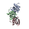
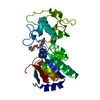
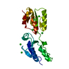
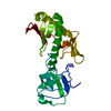
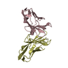
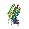
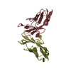
 Homo sapiens (human)
 X-RAY DIFFRACTION /
  Authors
 Citation
 Structure visualization
 Downloads & links
 Other downloads


 PDBj
PDBj


 Assembly


 Mass: 62.068 Da / Num. of mol.: 3 / Source method: obtained synthetically / Formula: C2H6O2
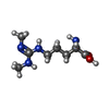
 Type: L-peptide linking / Mass: 202.254 Da / Num. of mol.: 1 / Source method: obtained synthetically / Formula: C8H18N4O2
 Mass: 18.015 Da / Num. of mol.: 79 / Source method: isolated from a natural source / Formula: H2O
 Sample preparation
 / Beamline: I04 / Wavelength: 1.0282 Å
 Processing