 Movie
Movie Controller

[English] 日本語
 Yorodumi
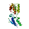
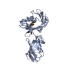
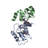
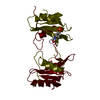
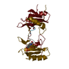
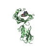
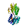
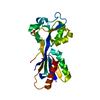
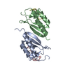
- PDB-1a2x: COMPLEX OF TROPONIN C WITH A 47 RESIDUE (1-47) FRAGMENT OF TROPONIN I -
+ Open data

- Basic information
| Entry | Database: PDB / ID: 1a2x | ||||||
|---|---|---|---|---|---|---|---|
| Title | COMPLEX OF TROPONIN C WITH A 47 RESIDUE (1-47) FRAGMENT OF TROPONIN I | ||||||
 Components Components |
| ||||||
 Keywords Keywords | COMPLEX (SKELETAL MUSCLE/MUSCLE PROTEIN) / TROPONIN / MUSCLE CONTRACTION REGULATION / COMPLEX (SKELETAL MUSCLE-MUSCLE PROTEIN) / COMPLEX (SKELETAL MUSCLE-MUSCLE PROTEIN) complex | ||||||
| Function / homology |  Function and homology information Function and homology informationtroponin T binding / troponin complex / myosin II complex / skeletal muscle contraction / cardiac muscle contraction / actin binding / calcium ion binding Similarity search - Function | ||||||
| Biological species |  | ||||||
| Method |  X-RAY DIFFRACTION / SYNCHROTRON / SIR/MAD / Resolution: 2.3 Å X-RAY DIFFRACTION / SYNCHROTRON / SIR/MAD / Resolution: 2.3 Å | ||||||
 Authors Authors | Vassylyev, D.G. / Takeda, S. / Wakatsuki, S. / Maeda, K. / Maeda, Y. | ||||||
 Citation Citation | Journal: Proc.Natl.Acad.Sci.USA / Year: 1998 Title: Crystal structure of troponin C in complex with troponin I fragment at 2.3-A resolution. Authors: Vassylyev, D.G. / Takeda, S. / Wakatsuki, S. / Maeda, K. / Maeda, Y. #1: Journal: Protein Sci. / Year: 1997Title: Production, Crystallization, and Preliminary X-Ray Analysis of Rabbit Skeletal Muscle Troponin Complex Consisting of Troponin C and Fragment (1-47) of Troponin I Authors: Saijo, Y. / Takeda, S. / Scherer, A. / Kobayashi, T. / Maeda, Y. / Taniguchi, H. / Yao, M. / Wakatsuki, S. | ||||||
| History |
|
- Structure visualization
| Structure viewer | Molecule: MolmilJmol/JSmol |
|---|
- Downloads & links
-Download
| PDBx/mmCIF format | 1a2x.cif.gz | 52.9 KB | Display | PDBx/mmCIF format |
|---|---|---|---|---|
| PDB format | pdb1a2x.ent.gz | 37.7 KB | Display | PDB format |
| PDBx/mmJSON format | 1a2x.json.gz | Tree view | PDBx/mmJSON format | |
| Others |  Other downloads Other downloads |
-Validation report
| Arichive directory | https://data.pdbj.org/pub/pdb/validation_reports/a2/1a2xftp://data.pdbj.org/pub/pdb/validation_reports/a2/1a2x | HTTPS FTP |
|---|
-Related structure data
| Similar structure data |
|---|

-Links
 PDBj
PDBj

- Assembly
| Deposited unit | 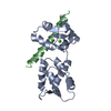
| ||||||||
|---|---|---|---|---|---|---|---|---|---|
| 1 |
| ||||||||
| Unit cell |
|
-Components
| #1: Protein | Mass: 17981.785 Da / Num. of mol.: 1 Source method: isolated from a genetically manipulated source Source: (gene. exp.)  | ||
|---|---|---|---|
| #2: Protein/peptide | Mass: 5546.197 Da / Num. of mol.: 1 / Fragment: RESIDUES 1 - 47 Source method: isolated from a genetically manipulated source Source: (gene. exp.) | ||
| #3: Chemical |   Mass: 40.078 Da / Num. of mol.: 2 / Source method: obtained synthetically / Formula: Ca Mass: 40.078 Da / Num. of mol.: 2 / Source method: obtained synthetically / Formula: Ca#4: Water | ChemComp-HOH / |  Mass: 18.015 Da / Num. of mol.: 89 / Source method: isolated from a natural source / Formula: H2O Mass: 18.015 Da / Num. of mol.: 89 / Source method: isolated from a natural source / Formula: H2O |
-Experimental details
-Experiment
| Experiment | Method: X-RAY DIFFRACTION / Number of used crystals: 1 |
|---|
- Sample preparation
| Crystal | Density Matthews: 2.3 Å3/Da / Density % sol: 35 % | ||||||||||||||||||||||||||||||
|---|---|---|---|---|---|---|---|---|---|---|---|---|---|---|---|---|---|---|---|---|---|---|---|---|---|---|---|---|---|---|---|
| Crystal grow | Temperature: 289 K / Method: vapor diffusion, hanging drop / pH: 8 Details: HANGING-DROP VAPOR DIFFUSION METHOD WAS USED AT 289K BY MIXING THE PROTEIN SOLUTION CONTAINING 25-30MG/ML OF THE CI47 COMPLEX WITH A RESERVOIR SOLUTION CONTAINING 1.5M SODIUM CITRATE, 0.1M ...Details: HANGING-DROP VAPOR DIFFUSION METHOD WAS USED AT 289K BY MIXING THE PROTEIN SOLUTION CONTAINING 25-30MG/ML OF THE CI47 COMPLEX WITH A RESERVOIR SOLUTION CONTAINING 1.5M SODIUM CITRATE, 0.1M TRIS-HCL, PH 8.0, 15% TREHALOSE., vapor diffusion - hanging drop | ||||||||||||||||||||||||||||||
| Crystal | *PLUS | ||||||||||||||||||||||||||||||
| Crystal grow | *PLUS Temperature: 16 ℃ / Method: vapor diffusion, hanging drop / Details: Saijo, Y., (1997) Protein Sci., 6, 916. | ||||||||||||||||||||||||||||||
| Components of the solutions | *PLUS
|
-Data collection
| Diffraction | Mean temperature: 100 K |
|---|---|
| Diffraction source | Source: SYNCHROTRON / Site: ESRF  / Beamline: BM14 / Wavelength: 0.97 / Beamline: BM14 / Wavelength: 0.97 |
| Detector | Type: MAR scanner 300 mm plate / Detector: IMAGE PLATE / Date: Jun 1, 1997 |
| Radiation | Monochromator: MONOCHROMATOR / Monochromatic (M) / Laue (L): M / Scattering type: x-ray |
| Radiation wavelength | Wavelength: 0.97 Å / Relative weight: 1 |
| Reflection | Resolution: 2.3→25 Å / Num. obs: 8870 / % possible obs: 96.3 % / Observed criterion σ(I): 0 / Redundancy: 4.5 % / Biso Wilson estimate: 45 Å2 / Rmerge(I) obs: 0.05 / Rsym value: 0.05 / Net I/σ(I): 9.2 |
| Reflection shell | Resolution: 2.3→2.4 Å / Redundancy: 4.3 % / Rmerge(I) obs: 0.3 / Mean I/σ(I) obs: 2.3 / % possible all: 94 |
| Reflection | *PLUS Num. measured all: 37214 |
- Processing
| Software |
| ||||||||||||||||||||||||||||||||||||||||||||||||||||||||||||||||||||||||||||||||
|---|---|---|---|---|---|---|---|---|---|---|---|---|---|---|---|---|---|---|---|---|---|---|---|---|---|---|---|---|---|---|---|---|---|---|---|---|---|---|---|---|---|---|---|---|---|---|---|---|---|---|---|---|---|---|---|---|---|---|---|---|---|---|---|---|---|---|---|---|---|---|---|---|---|---|---|---|---|---|---|---|---|
| Refinement | Method to determine structure: SIR/MAD / Resolution: 2.3→10 Å / Data cutoff high absF: 100000000000 / Data cutoff low absF: 0 / Isotropic thermal model: RESTRAINED / σ(F): 0
| ||||||||||||||||||||||||||||||||||||||||||||||||||||||||||||||||||||||||||||||||
| Displacement parameters | Biso mean: 41 Å2 | ||||||||||||||||||||||||||||||||||||||||||||||||||||||||||||||||||||||||||||||||
| Refine analyze | Luzzati coordinate error obs: 0.3 Å / Luzzati d res low obs: 6 Å / Luzzati sigma a obs: 0.28 Å | ||||||||||||||||||||||||||||||||||||||||||||||||||||||||||||||||||||||||||||||||
| Refinement step | Cycle: LAST / Resolution: 2.3→10 Å
| ||||||||||||||||||||||||||||||||||||||||||||||||||||||||||||||||||||||||||||||||
| Refine LS restraints |
| ||||||||||||||||||||||||||||||||||||||||||||||||||||||||||||||||||||||||||||||||
| LS refinement shell | Resolution: 2.3→2.4 Å / Total num. of bins used: 8
| ||||||||||||||||||||||||||||||||||||||||||||||||||||||||||||||||||||||||||||||||
| Xplor file |
| ||||||||||||||||||||||||||||||||||||||||||||||||||||||||||||||||||||||||||||||||
| Software | *PLUS Name: X-PLOR / Version: 3.1 / Classification: refinement | ||||||||||||||||||||||||||||||||||||||||||||||||||||||||||||||||||||||||||||||||
| Refine LS restraints | *PLUS
|
