 Movie
Movie Controller

[English] 日本語
 Yorodumi
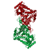
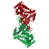
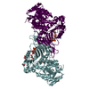
- PDB-1e7y: ACTIVE SITE MUTANT (D177->N) OF GLUCOSE 6-PHOSPHATE DEHYDROGENASE... -
+ Open data

- Basic information
| Entry | Database: PDB / ID: 1e7y | ||||||
|---|---|---|---|---|---|---|---|
| Title | ACTIVE SITE MUTANT (D177->N) OF GLUCOSE 6-PHOSPHATE DEHYDROGENASE FROM LEUCONOSTOC MESENTEROIDES COMPLEXED WITH SUBSTRATE AND NADPH | ||||||
 Components Components | GLUCOSE 6-PHOSPHATE 1-DEHYDROGENASE | ||||||
 Keywords Keywords | OXIDOREDUCTASE / OXIDOREDUCTASE (CHOH(D) - NAD(P)) / GLUCOSE METABOLISM | ||||||
| Function / homology |  Function and homology information Function and homology informationglucose-6-phosphate dehydrogenase [NAD(P)+] / glucose-6-phosphate dehydrogenase activity / pentose-phosphate shunt, oxidative branch / glucose metabolic process / NADP binding / cytosol Similarity search - Function | ||||||
| Biological species |  LEUCONOSTOC MESENTEROIDES (bacteria) LEUCONOSTOC MESENTEROIDES (bacteria) | ||||||
| Method |  X-RAY DIFFRACTION / MOLECULAR REPLACEMENT / Resolution: 2.48 Å X-RAY DIFFRACTION / MOLECULAR REPLACEMENT / Resolution: 2.48 Å | ||||||
 Authors Authors | Adams, M.J. / Cosgrove, M.S. / Gover, S. | ||||||
 Citation Citation | Journal: Biochemistry / Year: 2000 Title: An Examination of the Role of Asp-177 in the His-Asp Catalytic Dyad of Leuconostoc Mesenteroides Glucose 6-Phosphate Dehydrogenase: X-Ray Structure and Ph Dependence of Kinetic Parameters of ...Title: An Examination of the Role of Asp-177 in the His-Asp Catalytic Dyad of Leuconostoc Mesenteroides Glucose 6-Phosphate Dehydrogenase: X-Ray Structure and Ph Dependence of Kinetic Parameters of the D177N Mutant Enzyme Authors: Cosgrove, M.S. / Gover, S. / Naylor, C.E. / Vandeputte-Rutten, L. / Adams, M.J. / Levy, H.R. #1: Journal: Biochemistry / Year: 1998Title: On the Mechanism of the Reaction Catalysed by Glucose 6-Phosphate Dehydrogenase Authors: Cosgrove, M.S. / Naylor, C. / Paludin, S. / Adams, M.J. / Levy, H.R. #2: Journal: Structure / Year: 1994Title: The Three-Dimensional Structure of Glucose 6-Phosphate Dehydrogenase from Leuconostoc Mesenteroides Refined at 2 Angstroms Resolution Authors: Rowland, P. / Basak, A.K. / Gover, S. / Levy, H.R. / Adams, M.J. | ||||||
| History |
| ||||||
| Remark 650 | HELIX DETERMINATION METHOD: PROCHECK, WITH IDENTIFICATION CORRESPONDING TO 2.0A L. MESENTEROIDES ... HELIX DETERMINATION METHOD: PROCHECK, WITH IDENTIFICATION CORRESPONDING TO 2.0A L. MESENTEROIDES STRUCTURE, 1DPG. HELIX_ID: A,BEND AT K21 IS CONSEQUENCE OF CONSERVED P24. HELIX_ID: B,THE LAST TURN IS 3_10 (CLASS 5). HELIX_ID: C,THE FIRST TURN IS 3_10 (CLASS 5). HELIX_ID: D,THE FIRST TURN IS 3_10 (CLASS 5). HELIX_ID: F,THE FIRST TURN IS 3_10 (CLASS 5). HELIX_ID: H,G231 BRIDGES H & I' SO IS NOT HELICAL. HELIX_ID: I',PART OF HELIX I IN 1DPG. RESIDUES 235-239 DISTORTED BY SIDECHAIN INTERACTION OF N239 WITH D235. | ||||||
| Remark 700 | SHEET DETERMINATION METHOD: INITIAL AND TERMINAL RESIDUES ARE AS DEFINED BY PROCHECK. REGISTRATION ... SHEET DETERMINATION METHOD: INITIAL AND TERMINAL RESIDUES ARE AS DEFINED BY PROCHECK. REGISTRATION IS AS GIVEN BY HYDROGEN BONDS AND IN THE CASE OF SHEET COE INVOLVES RESIDUES THAT IMMEDIATELY PRECEDE EACH SHEET ELEMENT. THIS IS DONE TO PRESERVE OBSERVED CONSISTENCY WITH NATIVE STRUCTURE 1DPG. |
- Structure visualization
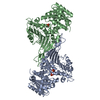
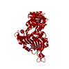
| Structure viewer | Molecule: MolmilJmol/JSmol |
|---|
- Downloads & links
-Download
| PDBx/mmCIF format | 1e7y.cif.gz | 113.7 KB | Display | PDBx/mmCIF format |
|---|---|---|---|---|
| PDB format | pdb1e7y.ent.gz | 86.6 KB | Display | PDB format |
| PDBx/mmJSON format | 1e7y.json.gz | Tree view | PDBx/mmJSON format | |
| Others |  Other downloads Other downloads |
-Validation report
| Arichive directory | https://data.pdbj.org/pub/pdb/validation_reports/e7/1e7yftp://data.pdbj.org/pub/pdb/validation_reports/e7/1e7y | HTTPS FTP |
|---|
-Related structure data


-Links
 PDBj
PDBj

- Assembly
| Deposited unit | 
| ||||||||
|---|---|---|---|---|---|---|---|---|---|
| 1 | 
| ||||||||
| Unit cell |
| ||||||||
| Components on special symmetry positions |
| ||||||||
| Details | BIOMOLECULE DIMERIC |
-Components
| #1: Protein | Mass: 54368.660 Da / Num. of mol.: 1 / Mutation: YES Source method: isolated from a genetically manipulated source Source: (gene. exp.) LEUCONOSTOC MESENTEROIDES (bacteria)Description: SITE DIRECTED MUTAGENESIS OTHER_DETAILS: SITE DIRECTED MUTAGENESIS Gene: PLMZ/D177NURCE 9 / Plasmid: PUC-19/ASP177ASNPLMZ / Gene (production host): PLMZ/D177N / Production host: References: UniProt: P11411, glucose-6-phosphate dehydrogenase (NADP+) |
|---|---|
| #2: Sugar | ChemComp-BG6 / 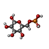  Type: D-saccharide, beta linking / Mass: 260.136 Da / Num. of mol.: 1 Type: D-saccharide, beta linking / Mass: 260.136 Da / Num. of mol.: 1Source method: isolated from a genetically manipulated source Formula: C6H13O9P |
| #3: Chemical | ChemComp-CA /   Mass: 40.078 Da / Num. of mol.: 1 / Source method: obtained synthetically / Formula: Ca Mass: 40.078 Da / Num. of mol.: 1 / Source method: obtained synthetically / Formula: Ca |
| #4: Chemical | ChemComp-NDP /   Mass: 745.421 Da / Num. of mol.: 1 / Source method: obtained synthetically / Formula: C21H30N7O17P3 Mass: 745.421 Da / Num. of mol.: 1 / Source method: obtained synthetically / Formula: C21H30N7O17P3 |
| #5: Water | ChemComp-HOH /  Mass: 18.015 Da / Num. of mol.: 131 / Source method: isolated from a natural source / Formula: H2O Mass: 18.015 Da / Num. of mol.: 131 / Source method: isolated from a natural source / Formula: H2O |
| Compound details | CHAIN A ENGINEERED MUTATION ASP177ASN BETA-D-GLUCOSE 6-PHOSPHATE + NADP(+) = D-GLUCONO-DELTA- ...CHAIN A ENGINEERED |
-Experimental details
-Experiment
| Experiment | Method: X-RAY DIFFRACTION / Number of used crystals: 1 |
|---|
- Sample preparation
| Crystal | Density Matthews: 2.3 Å3/Da / Density % sol: 42.2 % Description: MOLECULE LOCATED BY DIFFERENCE FOURIER AND RIGID-BODY REFINEMENT | ||||||||||||||||||||||||||||||||||||||||||
|---|---|---|---|---|---|---|---|---|---|---|---|---|---|---|---|---|---|---|---|---|---|---|---|---|---|---|---|---|---|---|---|---|---|---|---|---|---|---|---|---|---|---|---|
| Crystal grow | Method: vapor diffusion, hanging drop / pH: 7.5 Details: HANGING DROP VAPOUR DIFFUSION, 2+2 MICROLITER DROPS. IN THE WELL BUFFER: 14% W/V PEG 400 IN 0.1M HEPES-NAOH, PH 7.5 WITH 0.2M CALCIUM CHLORIDE. THE PROTEIN AT 15MG/ML, WITH 2.3MM GLUCOSE 6- ...Details: HANGING DROP VAPOUR DIFFUSION, 2+2 MICROLITER DROPS. IN THE WELL BUFFER: 14% W/V PEG 400 IN 0.1M HEPES-NAOH, PH 7.5 WITH 0.2M CALCIUM CHLORIDE. THE PROTEIN AT 15MG/ML, WITH 2.3MM GLUCOSE 6-PHOSPHATE AND 0.6MM NADPH. | ||||||||||||||||||||||||||||||||||||||||||
| Crystal grow | *PLUS Method: vapor diffusion, hanging drop | ||||||||||||||||||||||||||||||||||||||||||
| Components of the solutions | *PLUS
|
-Data collection
| Diffraction | Mean temperature: 100 K |
|---|---|
| Diffraction source | Source: ROTATING ANODE / Type: RIGAKU RUH2R / Wavelength: 1.5418 |
| Detector | Type: MARRESEARCH / Detector: IMAGE PLATE / Date: Jan 15, 1998 / Details: MIRRORS |
| Radiation | Monochromator: GRAPHITE(002) / Protocol: SINGLE WAVELENGTH / Monochromatic (M) / Laue (L): M / Scattering type: x-ray |
| Radiation wavelength | Wavelength: 1.5418 Å / Relative weight: 1 |
| Reflection | Resolution: 2.48→22.9 Å / Num. obs: 16723 / % possible obs: 93.6 % / Observed criterion σ(I): -3 / Redundancy: 2.6 % / Rsym value: 0.106 / Net I/σ(I): 9 |
| Reflection shell | Resolution: 2.48→2.59 Å / Redundancy: 2.2 % / Mean I/σ(I) obs: 2.7 / Rsym value: 0.46 / % possible all: 82.1 |
| Reflection | *PLUS Num. measured all: 38083 / Rmerge(I) obs: 0.106 |
| Reflection shell | *PLUS % possible obs: 82.1 % / Rmerge(I) obs: 0.46 |
- Processing
| Software |
| ||||||||||||||||||||||||||||||||||||||||||||||||||||||||||||||||||||||||||||||||
|---|---|---|---|---|---|---|---|---|---|---|---|---|---|---|---|---|---|---|---|---|---|---|---|---|---|---|---|---|---|---|---|---|---|---|---|---|---|---|---|---|---|---|---|---|---|---|---|---|---|---|---|---|---|---|---|---|---|---|---|---|---|---|---|---|---|---|---|---|---|---|---|---|---|---|---|---|---|---|---|---|---|
| Refinement | Method to determine structure: MOLECULAR REPLACEMENT Starting model: REFINED COORDINATES OF APO-ENZYME IN THE SAME SPACEGROUP Resolution: 2.48→22.9 Å / Data cutoff high absF: 100000 / Data cutoff low absF: 0 / Cross valid method: FREE R-VALUE / σ(F): 0 / Details: BULK SOLVENT WAS MODELLED.
| ||||||||||||||||||||||||||||||||||||||||||||||||||||||||||||||||||||||||||||||||
| Displacement parameters | Biso mean: 30.1 Å2 | ||||||||||||||||||||||||||||||||||||||||||||||||||||||||||||||||||||||||||||||||
| Refine analyze | Luzzati d res low obs: 22.9 Å / Luzzati sigma a obs: 0.52 Å | ||||||||||||||||||||||||||||||||||||||||||||||||||||||||||||||||||||||||||||||||
| Refinement step | Cycle: LAST / Resolution: 2.48→22.9 Å
| ||||||||||||||||||||||||||||||||||||||||||||||||||||||||||||||||||||||||||||||||
| Refine LS restraints |
| ||||||||||||||||||||||||||||||||||||||||||||||||||||||||||||||||||||||||||||||||
| LS refinement shell | Resolution: 2.48→2.59 Å / Total num. of bins used: 10
| ||||||||||||||||||||||||||||||||||||||||||||||||||||||||||||||||||||||||||||||||
| Xplor file |
| ||||||||||||||||||||||||||||||||||||||||||||||||||||||||||||||||||||||||||||||||
| Software | *PLUS Name: X-PLOR / Version: 3.851 / Classification: refinement | ||||||||||||||||||||||||||||||||||||||||||||||||||||||||||||||||||||||||||||||||
| Refinement | *PLUS Num. reflection obs: 15926 | ||||||||||||||||||||||||||||||||||||||||||||||||||||||||||||||||||||||||||||||||
| Solvent computation | *PLUS | ||||||||||||||||||||||||||||||||||||||||||||||||||||||||||||||||||||||||||||||||
| Displacement parameters | *PLUS | ||||||||||||||||||||||||||||||||||||||||||||||||||||||||||||||||||||||||||||||||
| Refine LS restraints | *PLUS
| ||||||||||||||||||||||||||||||||||||||||||||||||||||||||||||||||||||||||||||||||
| LS refinement shell | *PLUS Num. reflection Rwork: 1720 |
