 Movie
Movie Controller


+ Open data

- Basic information
| Entry | Database: PDB / ID: 1dpg | ||||||
|---|---|---|---|---|---|---|---|
| Title | GLUCOSE 6-PHOSPHATE DEHYDROGENASE FROM LEUCONOSTOC MESENTEROIDES | ||||||
 Components Components | GLUCOSE 6-PHOSPHATE DEHYDROGENASE | ||||||
 Keywords Keywords | OXIDOREDUCTASE (CHOH(D) - NAD(P)) / OXIDOREDUCTASE / NADP/NAD / GLUCOSE METABOLISM | ||||||
| Function / homology |  Function and homology information Function and homology informationglucose-6-phosphate dehydrogenase [NAD(P)+] / glucose-6-phosphate dehydrogenase activity / pentose-phosphate shunt, oxidative branch / glucose metabolic process / NADP binding / cytosol Similarity search - Function | ||||||
| Biological species |  Leuconostoc mesenteroides (bacteria) Leuconostoc mesenteroides (bacteria) | ||||||
| Method |  X-RAY DIFFRACTION / SYNCHROTRON / Resolution: 2 Å X-RAY DIFFRACTION / SYNCHROTRON / Resolution: 2 Å | ||||||
 Authors Authors | Adams, M.J. / Rowland, P. / Gover, S. | ||||||
 Citation Citation | Journal: Structure / Year: 1994 Title: The three-dimensional structure of glucose 6-phosphate dehydrogenase from Leuconostoc mesenteroides refined at 2.0 A resolution. Authors: Rowland, P. / Basak, A.K. / Gover, S. / Levy, H.R. / Adams, M.J. #1: Journal: Protein Sci. / Year: 1993Title: Site-Directed Mutagenesis to Facilitate X-Ray Structural Studies of Leuconostoc Mesenteroides Glucose 6-Phosphate Dehydrogenase Authors: Adams, M.J. / Basak, A.K. / Gover, S. / Rowland, P. / Levy, H.R. | ||||||
| History |
| ||||||
| Remark 650 | HELIX HELIX DETERMINATION METHOD: PROCHECK (I.E. DSSP) IN SUBUNIT A: HELIX_ID: A,BEND AT LYS 21 IS ...HELIX HELIX DETERMINATION METHOD: PROCHECK (I.E. DSSP) IN SUBUNIT A: HELIX_ID: A,BEND AT LYS 21 IS A CONSEQUENCE OF THE CONSERVED PRO 24. HELIX_ID: B,LAST TURN IS 3/10 (CLASS 5). HELIX_ID: D,FIRST TURN IS 3/10 (CLASS 5). HELIX_ID: F,FIRST 2 TURNS ARE 3/10 (CLASS 5). HELIX_ID: H,GLY 231 BRIDGES HELICES H AND I; IT IS NOT HELICAL. HELIX_ID: I,RESIDUES 235 - 239 ARE INFLUENCED BY AN ACTIVE SITE WATER MOLECULE. IN SUBUNIT B: HELIX_ID: A,BEND AT LYS 21 IS A CONSEQUENCE OF THE CONSERVED PRO 24. HELIX_ID: B,LAST TURN IS 3/10 (CLASS 5). HELIX_ID: D,FIRST TURN IS 3/10 (CLASS 5). HELIX_ID: F,FIRST TURN IS 3/10 (CLASS 5). HELIX_ID: H,GLY 231 BRIDGES HELICES H AND I; IT IS NOT HELICAL. HELIX_ID: I,RESIDUES 235 - 239 ARE INFLUENCED BY AN ACTIVE SITE WATER MOLECULE. | ||||||
| Remark 700 | SHEET SHEET SHEET_ID: COE; DETERMINATION METHOD: PROCHECK (I.E. DSSP, WITH EXTENSION TAKEN WHERE ...SHEET SHEET SHEET_ID: COE; DETERMINATION METHOD: PROCHECK (I.E. DSSP, WITH EXTENSION TAKEN WHERE HYDROGEN BONDING INDICATES THAT THIS IS APPROPRIATE). |
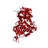
- Structure visualization
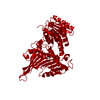
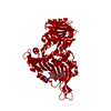
| Structure viewer | Molecule: MolmilJmol/JSmol |
|---|
- Downloads & links
-Download
| PDBx/mmCIF format | 1dpg.cif.gz | 212.7 KB | Display | PDBx/mmCIF format |
|---|---|---|---|---|
| PDB format | pdb1dpg.ent.gz | 170.4 KB | Display | PDB format |
| PDBx/mmJSON format | 1dpg.json.gz | Tree view | PDBx/mmJSON format | |
| Others |  Other downloads Other downloads |
-Validation report
| Arichive directory | https://data.pdbj.org/pub/pdb/validation_reports/dp/1dpgftp://data.pdbj.org/pub/pdb/validation_reports/dp/1dpg | HTTPS FTP |
|---|
-Related structure data
| Similar structure data |
|---|


-Links
 PDBj
PDBj- Assembly
| Deposited unit | 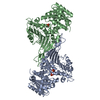
| ||||||||
|---|---|---|---|---|---|---|---|---|---|
| 1 |
| ||||||||
| Unit cell |
| ||||||||
| Atom site foot note | 1: CIS PROLINE - PRO A 149 / 2: CIS PROLINE - PRO A 375 / 3: CIS PROLINE - PRO B 375 | ||||||||
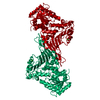
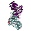
| Noncrystallographic symmetry (NCS) | NCS oper: (Code: given Matrix: (-0.3997, -0.7598, 0.5128), Vector: Details | MTRIX THE TRANSFORMATIONS PRESENTED ON MTRIX RECORDS BELOW DESCRIBE NON-CRYSTALLOGRAPHIC RELATIONSHIPS AMONG THE VARIOUS DOMAINS IN THIS ENTRY. THE ENZYME IS DIMERIC WITH A DIMER IN THE ASYMMETRIC UNIT. THE TRANSFORMATION GIVEN IS FOR BEST (LEAST SQUARES) SUPERPOSITION OF THE C-ALPHA ATOMS OF MONOMER B ONTO THOSE OF MONOMER A, SO APPLYING IT TO THE RESIDUES LISTED FIRST WILL YOELD APPROXIMATE COORDINATES FOR THE RESIDUES LISTED SECOND. THE ROTATION ANGLE IS 178.6 DEGREES AND THE AXIS IS AT 26.9 DEGREES TO THE AB PLANE. APPLIED TO TRANSFORMED TO MTRIX RESIDUES RESIDUES RMSD M1 B 1 .. B 485 A 1 .. A 485 0.724 | |
-Components
| #1: Protein | Mass: 54385.711 Da / Num. of mol.: 2 / Mutation: S61C Source method: isolated from a genetically manipulated source Source: (gene. exp.) Leuconostoc mesenteroides (bacteria) / Gene: G6PD / Plasmid: PLMZ / Gene (production host): G6PD / Production host: References: UniProt: P11411, glucose-6-phosphate dehydrogenase (NADP+) #2: Chemical |   Mass: 94.971 Da / Num. of mol.: 3 / Source method: obtained synthetically / Formula: PO4 Mass: 94.971 Da / Num. of mol.: 3 / Source method: obtained synthetically / Formula: PO4#3: Water | ChemComp-HOH / |  Mass: 18.015 Da / Num. of mol.: 610 / Source method: isolated from a natural source / Formula: H2O Mass: 18.015 Da / Num. of mol.: 610 / Source method: isolated from a natural source / Formula: H2O |
|---|
-Experimental details
-Experiment
| Experiment | Method: X-RAY DIFFRACTION |
|---|
- Sample preparation
| Crystal | Density Matthews: 3.33 Å3/Da / Density % sol: 63.01 % / Description: MERGE OF 7 DATA SETS (SEE JRNL) | ||||||||||||||||||||||||
|---|---|---|---|---|---|---|---|---|---|---|---|---|---|---|---|---|---|---|---|---|---|---|---|---|---|
| Crystal | *PLUS Density % sol: 64.7 % | ||||||||||||||||||||||||
| Crystal grow | *PLUS Temperature: 20 ℃ / pH: 5 / Method: vapor diffusion, hanging drop | ||||||||||||||||||||||||
| Components of the solutions | *PLUS
|
-Data collection
| Diffraction source | Source: SYNCHROTRON / Site: SRS  / Beamline: PX9.5 / Wavelength: 0.88, 1.00 / Beamline: PX9.5 / Wavelength: 0.88, 1.00 | |||||||||
|---|---|---|---|---|---|---|---|---|---|---|
| Detector | Type: MARRESEARCH / Detector: IMAGE PLATE | |||||||||
| Radiation | Monochromatic (M) / Laue (L): M / Scattering type: x-ray | |||||||||
| Radiation wavelength |
| |||||||||
| Reflection | Resolution: 2→27 Å / Num. obs: 92804 / % possible obs: 94 % / Redundancy: 7.5 % | |||||||||
| Reflection | *PLUS Redundancy: 7.5 % |
- Processing
| Software |
| ||||||||||||||||||||||||||||||||||||||||||||||||||||||||||||
|---|---|---|---|---|---|---|---|---|---|---|---|---|---|---|---|---|---|---|---|---|---|---|---|---|---|---|---|---|---|---|---|---|---|---|---|---|---|---|---|---|---|---|---|---|---|---|---|---|---|---|---|---|---|---|---|---|---|---|---|---|---|
| Refinement | Resolution: 2→27 Å / σ(F): 0
| ||||||||||||||||||||||||||||||||||||||||||||||||||||||||||||
| Refine analyze | Luzzati coordinate error obs: 0.25 Å | ||||||||||||||||||||||||||||||||||||||||||||||||||||||||||||
| Refinement step | Cycle: LAST / Resolution: 2→27 Å
| ||||||||||||||||||||||||||||||||||||||||||||||||||||||||||||
| Refine LS restraints |
| ||||||||||||||||||||||||||||||||||||||||||||||||||||||||||||
| Software | *PLUS Name: X-PLOR / Classification: refinement | ||||||||||||||||||||||||||||||||||||||||||||||||||||||||||||
| Refinement | *PLUS | ||||||||||||||||||||||||||||||||||||||||||||||||||||||||||||
| Solvent computation | *PLUS | ||||||||||||||||||||||||||||||||||||||||||||||||||||||||||||
| Displacement parameters | *PLUS | ||||||||||||||||||||||||||||||||||||||||||||||||||||||||||||
| Refine LS restraints | *PLUS
|
