 Movie
Movie Controller

[English] 日本語
 Yorodumi
- PDB-2dpg: COMPLEX OF INACTIVE MUTANT (H240->N) OF GLUCOSE 6-PHOSPHATE DEHYD... -
+ Open data

- Basic information
| Entry | Database: PDB / ID: 2dpg | |||||||||
|---|---|---|---|---|---|---|---|---|---|---|
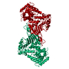
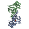
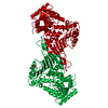
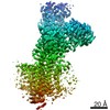
| Title | COMPLEX OF INACTIVE MUTANT (H240->N) OF GLUCOSE 6-PHOSPHATE DEHYDROGENASE FROM LEUCONOSTOC MESENTEROIDES WITH NADP+ | |||||||||
 Components Components | GLUCOSE 6-PHOSPHATE DEHYDROGENASE | |||||||||
 Keywords Keywords | OXIDOREDUCTASE / CHOH(D) - NAD(P) / NADP/NAD / GLUCOSE METABOLISM | |||||||||
| Function / homology |  Function and homology information Function and homology informationglucose-6-phosphate dehydrogenase [NAD(P)+] / glucose-6-phosphate dehydrogenase activity / pentose-phosphate shunt, oxidative branch / glucose metabolic process / NADP binding / cytosol Similarity search - Function | |||||||||
| Biological species |  Leuconostoc mesenteroides (bacteria) Leuconostoc mesenteroides (bacteria) | |||||||||
| Method |  X-RAY DIFFRACTION / SYNCHROTRON / RIGID-BODY, DIFFERENCE FOURIER / Resolution: 2.5 Å X-RAY DIFFRACTION / SYNCHROTRON / RIGID-BODY, DIFFERENCE FOURIER / Resolution: 2.5 Å | |||||||||
 Authors Authors | Adams, M.J. / Naylor, C.E. / Paludin, S. / Gover, S. | |||||||||
 Citation Citation | Journal: Biochemistry / Year: 1998 Title: On the mechanism of the reaction catalyzed by glucose 6-phosphate dehydrogenase. Authors: Cosgrove, M.S. / Naylor, C. / Paludan, S. / Adams, M.J. / Levy, H.R. #1: Journal: Structure / Year: 1994Title: The Three-Dimensional Structure of Glucose 6-Phosphate Dehydrogenase from Leuconostoc Mesenteroides Refined at 2.0 A Resolution Authors: Rowland, P. / Basak, A.K. / Gover, S. / Levy, H.R. / Adams, M.J. #2: Journal: Protein Sci. / Year: 1993Title: Site-Directed Mutagenesis to Facilitate X-Ray Structural Studies of Leuconostoc Mesenteroides Glucose 6-Phosphate Dehydrogenase Authors: Adams, M.J. / Basak, A.K. / Gover, S. / Rowland, P. / Levy, H.R. | |||||||||
| History |
|
- Structure visualization
| Structure viewer | Molecule: MolmilJmol/JSmol |
|---|
- Downloads & links
-Download
| PDBx/mmCIF format | 2dpg.cif.gz | 109.4 KB | Display | PDBx/mmCIF format |
|---|---|---|---|---|
| PDB format | pdb2dpg.ent.gz | 84.6 KB | Display | PDB format |
| PDBx/mmJSON format | 2dpg.json.gz | Tree view | PDBx/mmJSON format | |
| Others |  Other downloads Other downloads |
-Validation report
| Arichive directory | https://data.pdbj.org/pub/pdb/validation_reports/dp/2dpgftp://data.pdbj.org/pub/pdb/validation_reports/dp/2dpg | HTTPS FTP |
|---|
-Related structure data
| Similar structure data |
|---|


-Links
 PDBj
PDBj- Assembly
| Deposited unit | 
| ||||||||
|---|---|---|---|---|---|---|---|---|---|
| 1 | 
| ||||||||
| Unit cell |
| ||||||||
| Components on special symmetry positions |
|
-Components
| #1: Protein | Mass: 54345.602 Da / Num. of mol.: 1 / Mutation: H240N Source method: isolated from a genetically manipulated source Details: THE MUTANT IS INACTIVE, LACKING THE CATALYTIC BASE. THE STRUCTURE CONTAINS A PARTIAL NADP BOUND TO NUCLEOTIDE BINDING DOMAIN. Source: (gene. exp.) Leuconostoc mesenteroides (bacteria) / Gene: PLMZ/H240N / Plasmid: PUC-19/HIS240ASNPLMZ / Gene (production host): PLMZ/H240N / Production host: References: UniProt: P11411, glucose-6-phosphate dehydrogenase (NADP+) |
|---|---|
| #2: Chemical | ChemComp-NAP /   Mass: 743.405 Da / Num. of mol.: 1 / Source method: obtained synthetically / Formula: C21H28N7O17P3 Mass: 743.405 Da / Num. of mol.: 1 / Source method: obtained synthetically / Formula: C21H28N7O17P3 |
| #3: Water | ChemComp-HOH /  Mass: 18.015 Da / Num. of mol.: 74 / Source method: isolated from a natural source / Formula: H2O Mass: 18.015 Da / Num. of mol.: 74 / Source method: isolated from a natural source / Formula: H2O |
-Experimental details
-Experiment
| Experiment | Method: X-RAY DIFFRACTION / Number of used crystals: 2 |
|---|
- Sample preparation
| Crystal | Density Matthews: 2.98 Å3/Da / Density % sol: 60 % Description: MERGE OF 2.5 ANGSTROM SRS DATA (EXPT 1) WITH 3.5 ANGSTROM IN-HOUSE DATA (EXPT 2). FOR DETAILS, SEE JRNL REFERENCE. | |||||||||||||||||||||||||
|---|---|---|---|---|---|---|---|---|---|---|---|---|---|---|---|---|---|---|---|---|---|---|---|---|---|---|
| Crystal grow | Method: vapor diffusion, hanging drop / pH: 7.5 Details: HANGING DROP VAPOR DIFFUSION. 2+2 MICROLITER DROPS. IN THE WELL 2.27M UNBUFFERED AMMONIUM SULFATE. THE PROTEIN AT 4.8MG/ML IN 0.1M TRIS-HCL AT PH 7.5 WITH 0.5MM NADP+ AND 25MM G6P., vapor ...Details: HANGING DROP VAPOR DIFFUSION. 2+2 MICROLITER DROPS. IN THE WELL 2.27M UNBUFFERED AMMONIUM SULFATE. THE PROTEIN AT 4.8MG/ML IN 0.1M TRIS-HCL AT PH 7.5 WITH 0.5MM NADP+ AND 25MM G6P., vapor diffusion - hanging drop | |||||||||||||||||||||||||
| Crystal | *PLUS | |||||||||||||||||||||||||
| Crystal grow | *PLUS Method: vapor diffusion, hanging drop | |||||||||||||||||||||||||
| Components of the solutions | *PLUS
|
-Data collection
| Diffraction | Mean temperature: 293 K |
|---|---|
| Diffraction source | Source: SYNCHROTRON / Site: SRS  / Beamline: PX9.5 / Wavelength: 0.876 / Beamline: PX9.5 / Wavelength: 0.876 |
| Detector | Type: MARRESEARCH / Detector: IMAGE PLATE / Date: Dec 1, 1994 / Details: MIRRORS |
| Radiation | Monochromator: SI(111) / Monochromatic (M) / Laue (L): M / Scattering type: x-ray |
| Radiation wavelength | Wavelength: 0.876 Å / Relative weight: 1 |
| Reflection | Resolution: 2.5→30 Å / Num. obs: 16263 / % possible obs: 68.4 % / Observed criterion σ(I): -3 / Redundancy: 1.3 % / Biso Wilson estimate: 41.3 Å2 / Rmerge(I) obs: 0.08 / Net I/σ(I): 7.1 |
| Reflection shell | Resolution: 2.5→2.6 Å / Mean I/σ(I) obs: 2.2 / Rsym value: 0.411 / % possible all: 0.548 |
| Reflection | *PLUS Num. measured all: 21142 |
| Reflection shell | *PLUS Rmerge(I) obs: 0.125 |
- Processing
| Software |
| ||||||||||||||||||||||||||||||||||||||||||||||||||||||||||||||||||||||||||||||||
|---|---|---|---|---|---|---|---|---|---|---|---|---|---|---|---|---|---|---|---|---|---|---|---|---|---|---|---|---|---|---|---|---|---|---|---|---|---|---|---|---|---|---|---|---|---|---|---|---|---|---|---|---|---|---|---|---|---|---|---|---|---|---|---|---|---|---|---|---|---|---|---|---|---|---|---|---|---|---|---|---|---|
| Refinement | Method to determine structure: RIGID-BODY, DIFFERENCE FOURIER Starting model: INCOMPLETE REFINED STRUCTURE OF NATIVE PROTEIN CONTAINING NADP Resolution: 2.5→25 Å / Data cutoff high absF: 100000 / Data cutoff low absF: 0 / Cross valid method: FREE R / σ(F): 0 Details: FOR DETAILS OF THE BULK SOLVENT MODELLING, SEE JRNL REFERENCE.
| ||||||||||||||||||||||||||||||||||||||||||||||||||||||||||||||||||||||||||||||||
| Displacement parameters | Biso mean: 30.8 Å2 | ||||||||||||||||||||||||||||||||||||||||||||||||||||||||||||||||||||||||||||||||
| Refine analyze | Luzzati d res low obs: 25 Å / Luzzati sigma a obs: 0.3 Å | ||||||||||||||||||||||||||||||||||||||||||||||||||||||||||||||||||||||||||||||||
| Refinement step | Cycle: LAST / Resolution: 2.5→25 Å
| ||||||||||||||||||||||||||||||||||||||||||||||||||||||||||||||||||||||||||||||||
| Refine LS restraints |
| ||||||||||||||||||||||||||||||||||||||||||||||||||||||||||||||||||||||||||||||||
| LS refinement shell | Resolution: 2.5→2.59 Å / Total num. of bins used: 10
| ||||||||||||||||||||||||||||||||||||||||||||||||||||||||||||||||||||||||||||||||
| Xplor file |
| ||||||||||||||||||||||||||||||||||||||||||||||||||||||||||||||||||||||||||||||||
| Software | *PLUS Name: X-PLOR / Version: 3.1 / Classification: refinement | ||||||||||||||||||||||||||||||||||||||||||||||||||||||||||||||||||||||||||||||||
| Refine LS restraints | *PLUS
| ||||||||||||||||||||||||||||||||||||||||||||||||||||||||||||||||||||||||||||||||
| LS refinement shell | *PLUS Rfactor Rfree: 0.35 |
