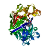
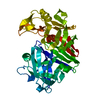
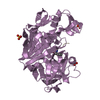
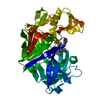
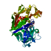
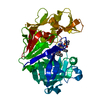
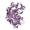
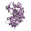
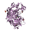
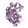
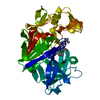
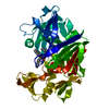
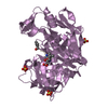
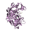
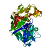
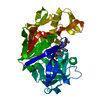
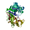
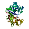
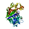
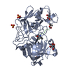
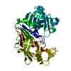
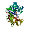
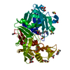
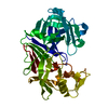
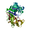
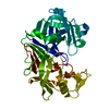
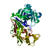
Journal: Thesis / Year: 1999 Title: Refinement of Four Endothiapepsin Inhibitor Complexes. Crystallographic Studies of Cytochrome Ch from Methylobacterium Extorquens and Inhibitor Complexes of Aspartic Proteinases. Authors: Read, J.A.
History
Deposition
Jul 28, 2000
Deposition site: PDBE / Processing site: PDBE
Revision 1.0
Sep 7, 2000
Provider: repository / Type: Initial release
Revision 1.1
Jul 13, 2011
Group: Atomic model / Database references ...Atomic model / Database references / Derived calculations / Non-polymer description / Structure summary / Version format compliance
Mass: 18.015 Da / Num. of mol.: 189 / Source method: isolated from a natural source / Formula: H2O
Compound details
CATALYTIC ACTIVITY: HYDROLYSIS OF PROTEINS WITH BROAD SPECIFICITY SIMILAR TO THAT OF PEPSIN A, ...CATALYTIC ACTIVITY: HYDROLYSIS OF PROTEINS WITH BROAD SPECIFICITY SIMILAR TO THAT OF PEPSIN A, PREFERRING HYDROPHOBIC RESIDUES AT P1 AND P1', BUT DOES NOT CLEAVE 14-ALA-|-LEU-15 IN THE B CHAIN OF INSULIN OR Z-GLU-TYR. CLOTS MILK. SIMILARITY: BELONGS TO PEPTIDASE FAMILY A1; ALSO KNOWN AS THE EUKARYOTIC ASPARTYL PROTEASES FAMILY.
Has protein modification
Y
-
Experimental details
-
Experiment
Experiment
Method: X-RAY DIFFRACTION / Number of used crystals: 1
-
Sample preparation
Crystal
Density Matthews: 2.48 Å3/Da / Density % sol: 50.5 %
Crystal grow
pH: 5 Details: BATCH METHOD PROTEIN: 10MG/ML, 50 MM AMMONIUM ACETATE PH 5.0, ~2.2 M AMMONIUM SULPHATE, 1% ACETONE. INHIBITOR WAS ADDED IN A 10:1 STOICHIOMETRIC RATIO.
-
Data collection
Diffraction
Mean temperature: 298 K
Diffraction source
Source: ROTATING ANODE / Wavelength: 1.5418
Detector
Type: MARRESEARCH / Detector: IMAGE PLATE
Radiation
Monochromator: NI FILTER / Protocol: SINGLE WAVELENGTH / Monochromatic (M) / Laue (L): M / Scattering type: x-ray
Radiation wavelength
Wavelength: 1.5418 Å / Relative weight: 1
Reflection
Resolution: 2.05→30 Å / Num. obs: 21510 / % possible obs: 98.9 % / Observed criterion σ(I): 0 / Redundancy: 3.8 % / Rsym value: 0.123 / Net I/σ(I): 5.3
Reflection shell
Resolution: 2.05→2.29 Å / Redundancy: 3.7 % / Mean I/σ(I) obs: 1.6 / Rsym value: 0.461 / % possible all: 94.3
In the structure databanks used in Yorodumi, some data are registered as the other names, "COVID-19 virus" and "2019-nCoV". Here are the details of the virus and the list of structure data.
Jan 31, 2019. EMDB accession codes are about to change! (news from PDBe EMDB page)
EMDB accession codes are about to change! (news from PDBe EMDB page)
The allocation of 4 digits for EMDB accession codes will soon come to an end. Whilst these codes will remain in use, new EMDB accession codes will include an additional digit and will expand incrementally as the available range of codes is exhausted. The current 4-digit format prefixed with “EMD-” (i.e. EMD-XXXX) will advance to a 5-digit format (i.e. EMD-XXXXX), and so on. It is currently estimated that the 4-digit codes will be depleted around Spring 2019, at which point the 5-digit format will come into force.
The EM Navigator/Yorodumi systems omit the EMD- prefix.
Related info.:Q: What is EMD? / ID/Accession-code notation in Yorodumi/EM Navigator
Yorodumi is a browser for structure data from EMDB, PDB, SASBDB, etc.
This page is also the successor to EM Navigator detail page, and also detail information page/front-end page for Omokage search.
The word "yorodu" (or yorozu) is an old Japanese word meaning "ten thousand". "mi" (miru) is to see.
Related info.:EMDB / PDB / SASBDB / Comparison of 3 databanks / Yorodumi Search / Aug 31, 2016. New EM Navigator & Yorodumi / Yorodumi Papers / Jmol/JSmol / Function and homology information / Changes in new EM Navigator and Yorodumi
 Movie
Movie Controller


 Open data
 Basic information
 Components
 Keywords
 Function and homology information
 ENDOTHIA PARASITICA (chestnut blight fungus)
 X-RAY DIFFRACTION /
  Authors
 Citation
 Structure visualization
 Downloads & links
 Other downloads


 PDBj
PDBj
 Assembly
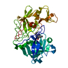
 Type: peptide-like / Mass: 466.637 Da / Num. of mol.: 1 / Source method: obtained synthetically / Formula: C23H38N4O4S
 Mass: 18.015 Da / Num. of mol.: 189 / Source method: isolated from a natural source / Formula: H2O
 Sample preparation
 Processing