 Movie
Movie Controller

[English] 日本語
 Yorodumi
- PDB-1e5n: E246C mutant of P fluorescens subsp. cellulosa xylanase A in comp... -
+ Open data

- Basic information
| Entry | Database: PDB / ID: 1e5n | |||||||||
|---|---|---|---|---|---|---|---|---|---|---|
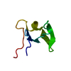
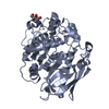
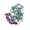
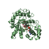
| Title | E246C mutant of P fluorescens subsp. cellulosa xylanase A in complex with xylopentaose | |||||||||
 Components Components | ENDO-1,4-BETA-XYLANASE A | |||||||||
 Keywords Keywords | HYDROLASE / GLYCOSYL HYDROLASE / FAMILY 10 / XYLAN DEGRADATION | |||||||||
| Function / homology |  Function and homology information Function and homology informationcellulose binding / endo-1,4-beta-xylanase / endo-1,4-beta-xylanase activity / xylan catabolic process Similarity search - Function | |||||||||
| Biological species |  PSEUDOMONAS FLUORESCENS (bacteria) PSEUDOMONAS FLUORESCENS (bacteria) | |||||||||
| Method |  X-RAY DIFFRACTION / FOURIER SYNTHESIS / Resolution: 3.2 Å X-RAY DIFFRACTION / FOURIER SYNTHESIS / Resolution: 3.2 Å | |||||||||
 Authors Authors | Lo Leggio, L. / Jenkins, J.A. / Harris, G.W. / Pickersgill, R.W. | |||||||||
 Citation Citation | Journal: Proteins / Year: 2000 Title: X-ray crystallographic study of xylopentaose binding to Pseudomonas fluorescens xylanase A. Authors: Leggio, L.L. / Jenkins, J. / Harris, G.W. / Pickersgill, R.W. #1: Journal: Enzyme Microb.Technol. / Year: 1999Title: Xylanase-Oligosaccharide Interactions Studied by a Competitive Enzyme Assay Authors: Lo Leggio, L. / Pickersgill, R.W. #2: Journal: Acta Crystallogr.,Sect.D / Year: 1996Title: Refined Crystal Structure of the Catalytic Domain of Xylanase a from Pseudomonas Fluorescens at 1.8 Angstrom Resolution Authors: Harris, G.W. / Jenkins, J.A. / Connerton, I. / Pickersgill, R.W. #3: Journal: Structure / Year: 1994Title: Structure of the Catalytic Core of the Family F Xylanase from Pseudomonas Fluorescens and Identification of the Xylopentaose-Binding Sites Authors: Harris, G.W. / Jenkins, J.A. / Connerton, I. / Cummings, N. / Lo Leggio, L. / Scott, M. / Hazlewood, G.P. / Laurie, J.I. / Gilbert, H.J. / Pickersgill, R.W. | |||||||||
| History |
|
- Structure visualization
| Structure viewer | Molecule: MolmilJmol/JSmol |
|---|
- Downloads & links
-Download
| PDBx/mmCIF format | 1e5n.cif.gz | 134.2 KB | Display | PDBx/mmCIF format |
|---|---|---|---|---|
| PDB format | pdb1e5n.ent.gz | 107.8 KB | Display | PDB format |
| PDBx/mmJSON format | 1e5n.json.gz | Tree view | PDBx/mmJSON format | |
| Others |  Other downloads Other downloads |
-Validation report
| Arichive directory | https://data.pdbj.org/pub/pdb/validation_reports/e5/1e5nftp://data.pdbj.org/pub/pdb/validation_reports/e5/1e5n | HTTPS FTP |
|---|
-Related structure data
| Related structure data |  1clxS S: Starting model for refinement |
|---|---|
| Similar structure data |


-Links
 PDBj
PDBj

- Assembly
| Deposited unit | 
| ||||||||
|---|---|---|---|---|---|---|---|---|---|
| 1 | 
| ||||||||
| 2 | 
| ||||||||
| Unit cell |
| ||||||||
| Noncrystallographic symmetry (NCS) | NCS oper: (Code: given Matrix: (-0.997297, 0.059884, 0.042586), Vector: |
-Components
| #1: Protein | Mass: 38548.609 Da / Num. of mol.: 2 / Fragment: CATALYTIC DOMAIN RESIDUES 264-611 / Mutation: YES Source method: isolated from a genetically manipulated source Details: TRUNCATED CATALYTIC DOMAIN, AA 264-611 / Source: (gene. exp.) PSEUDOMONAS FLUORESCENS (bacteria) / Variant: SUBSP CELLULOSA / Production host: #2: Polysaccharide | Source method: isolated from a genetically manipulated source #3: Chemical |   Mass: 40.078 Da / Num. of mol.: 2 / Source method: obtained synthetically / Formula: Ca Mass: 40.078 Da / Num. of mol.: 2 / Source method: obtained synthetically / Formula: CaCompound details | CHAIN A ENGINEERED | Has protein modification | Y | |
|---|
-Experimental details
-Experiment
| Experiment | Method: X-RAY DIFFRACTION / Number of used crystals: 1 |
|---|
- Sample preparation
| Crystal | Density Matthews: 2.32 Å3/Da / Density % sol: 46.87 % | ||||||||||||||||||||||||||||||
|---|---|---|---|---|---|---|---|---|---|---|---|---|---|---|---|---|---|---|---|---|---|---|---|---|---|---|---|---|---|---|---|
| Crystal grow | Method: vapor diffusion, hanging drop / pH: 6.5 Details: HANGING DROP (10 MG/ML OF PROTEIN) WITH A RESERVOIR OF 0.1 M SODIUM CACODYLATE PH 6.5, 200 MM CALCIUM ACETATE, 1 MM BETA-MERCAPTOETHANOL, 14-18% PEG 8000 | ||||||||||||||||||||||||||||||
| Crystal grow | *PLUS Method: vapor diffusion, hanging dropDetails: drop consists of equal amounts of protein and reservoir solutions | ||||||||||||||||||||||||||||||
| Components of the solutions | *PLUS
|
-Data collection
| Diffraction | Mean temperature: 293 K |
|---|---|
| Diffraction source | Source: ROTATING ANODE / Type: SIEMENS / Wavelength: 1.5418 |
| Detector | Type: MULTIWIRE SIEMENS / Detector: AREA DETECTOR |
| Radiation | Protocol: SINGLE WAVELENGTH / Monochromatic (M) / Laue (L): M / Scattering type: x-ray |
| Radiation wavelength | Wavelength: 1.5418 Å / Relative weight: 1 |
| Reflection | Resolution: 3.2→29.7 Å / Num. obs: 11399 / % possible obs: 98.9 % / Redundancy: 4.3 % / Rsym value: 0.144 |
| Reflection shell | Resolution: 3.2→3.4 Å / Redundancy: 3.1 % / Rsym value: 0.364 / % possible all: 95.8 |
| Reflection | *PLUS Rmerge(I) obs: 0.144 |
| Reflection shell | *PLUS % possible obs: 95.8 % / Rmerge(I) obs: 0.364 |
- Processing
| Software |
| ||||||||||||||||||||||||||||||||||||||||||||||||||||||||||||
|---|---|---|---|---|---|---|---|---|---|---|---|---|---|---|---|---|---|---|---|---|---|---|---|---|---|---|---|---|---|---|---|---|---|---|---|---|---|---|---|---|---|---|---|---|---|---|---|---|---|---|---|---|---|---|---|---|---|---|---|---|---|
| Refinement | Method to determine structure: FOURIER SYNTHESIS Starting model: PDB ENTRY 1CLX Resolution: 3.2→29.7 Å / Rfactor Rfree error: 0.0088 / Data cutoff high absF: 100000 / Data cutoff low absF: 0.1 / Isotropic thermal model: RESTRAINED GROUPED B FACTORS / Cross valid method: THROUGHOUT / σ(F): 0 Details: THE C-TERMINAL RESIDUE (ARG 347) WAS NOT VISIBLE IN THE ELECTRON DENSITY MAP
| ||||||||||||||||||||||||||||||||||||||||||||||||||||||||||||
| Refinement step | Cycle: LAST / Resolution: 3.2→29.7 Å
| ||||||||||||||||||||||||||||||||||||||||||||||||||||||||||||
| Refine LS restraints |
| ||||||||||||||||||||||||||||||||||||||||||||||||||||||||||||
| Xplor file |
| ||||||||||||||||||||||||||||||||||||||||||||||||||||||||||||
| Software | *PLUS Name: X-PLOR / Version: 3.1 / Classification: refinement | ||||||||||||||||||||||||||||||||||||||||||||||||||||||||||||
| Refine LS restraints | *PLUS
|
