 Movie
Movie Controller


+ Open data

- Basic information
| Entry | Database: PDB / ID: 1clx | ||||||
|---|---|---|---|---|---|---|---|
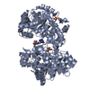
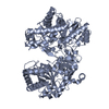
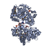
| Title | CATALYTIC CORE OF XYLANASE A | ||||||
 Components Components | XYLANASE A | ||||||
 Keywords Keywords | XYLANASE / FAMILY-F XYLANASE FAMILY 10 GLYCOSYL-HYDROLASE | ||||||
| Function / homology |  Function and homology information Function and homology informationcellulose binding / endo-1,4-beta-xylanase / endo-1,4-beta-xylanase activity / xylan catabolic process Similarity search - Function | ||||||
| Biological species |  Cellvibrio japonicus (bacteria) Cellvibrio japonicus (bacteria) | ||||||
| Method |  X-RAY DIFFRACTION / SYNCHROTRON / Resolution: 1.8 Å X-RAY DIFFRACTION / SYNCHROTRON / Resolution: 1.8 Å | ||||||
 Authors Authors | Harris, G.W. / Jenkins, J.A. / Connerton, I. / Pickersgill, R.W. | ||||||
 Citation Citation | Journal: Acta Crystallogr.,Sect.D / Year: 1996 Title: Refined crystal structure of the catalytic domain of xylanase A from Pseudomonas fluorescens at 1.8 A resolution. Authors: Harris, G.W. / Jenkins, J.A. / Connerton, I. / Pickersgill, R.W. #1: Journal: FEBS Lett. / Year: 1995Title: Beta-Glucosidase, Beta-Galactosidase, Family a Cellulases, Family F Xylanases and Two Barley Glycanases Form a Superfamily of Enzymes with 8-Fold Beta/Alpha Architecture and with Two Conserved ...Title: Beta-Glucosidase, Beta-Galactosidase, Family a Cellulases, Family F Xylanases and Two Barley Glycanases Form a Superfamily of Enzymes with 8-Fold Beta/Alpha Architecture and with Two Conserved Glutamates Near the Carboxy-Terminal Ends of Beta-Strands Four and Seven Authors: Jenkins, J. / Lo Leggio, L. / Harris, G. / Pickersgill, R. #2: Journal: Structure / Year: 1994Title: Structure of the Catalytic Core of the Family F Xylanase from Pseudomonas Fluorescens and Identification of the Xylopentaose-Binding Sites Authors: Harris, G.W. / Jenkins, J.A. / Connerton, I. / Cummings, N. / Lo Leggio, L. / Scott, M. / Hazlewood, G.P. / Laurie, J.I. / Gilbert, H.J. / Pickersgill, R.W. #3: Journal: J.Mol.Biol. / Year: 1993Title: Crystallization and Preliminary X-Ray Analysis of the Catalytic Domain of Xylanase a from Pseudomonas Fluorescens Subspecies Cellulosa Authors: Pickersgill, R.W. / Jenkins, J.A. / Scott, M. / Connerton, I. / Hazlewood, G.P. / Gilbert, H.J. | ||||||
| History |
|
- Structure visualization
| Structure viewer | Molecule: MolmilJmol/JSmol |
|---|
- Downloads & links
-Download
| PDBx/mmCIF format | 1clx.cif.gz | 296.4 KB | Display | PDBx/mmCIF format |
|---|---|---|---|---|
| PDB format | pdb1clx.ent.gz | 236.5 KB | Display | PDB format |
| PDBx/mmJSON format | 1clx.json.gz | Tree view | PDBx/mmJSON format | |
| Others |  Other downloads Other downloads |
-Validation report
| Arichive directory | https://data.pdbj.org/pub/pdb/validation_reports/cl/1clxftp://data.pdbj.org/pub/pdb/validation_reports/cl/1clx | HTTPS FTP |
|---|
-Related structure data
| Similar structure data |
|---|


-Links
 PDBj
PDBj

- Assembly
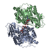
| Deposited unit | 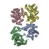
| ||||||||
|---|---|---|---|---|---|---|---|---|---|
| 1 | 
| ||||||||
| 2 | 
| ||||||||
| Unit cell |
|
-Components
| #1: Protein | Mass: 38460.477 Da / Num. of mol.: 4 / Fragment: CATALYTIC CORE, RESIDUES 264 - 611 Source method: isolated from a genetically manipulated source Source: (gene. exp.) Cellvibrio japonicus (bacteria) / Strain: CELLULOSA / Gene: TRUNCATED XYNA (CODONS 264-611 / Plasmid: PET3A / Gene (production host): TRUNCATED XYNA (CODONS 264-611) / Production host: #2: Chemical | ChemComp-CA /   Mass: 40.078 Da / Num. of mol.: 4 / Source method: obtained synthetically / Formula: Ca Mass: 40.078 Da / Num. of mol.: 4 / Source method: obtained synthetically / Formula: Ca#3: Water | ChemComp-HOH / |  Mass: 18.015 Da / Num. of mol.: 1108 / Source method: isolated from a natural source / Formula: H2O Mass: 18.015 Da / Num. of mol.: 1108 / Source method: isolated from a natural source / Formula: H2OCompound details | THE STRUCTURE IS OF THE CATALYTIC CORE OF XYLANASE A; THE COMPLETE XYLANASE WOULD ALSO CONSIST OF A ...THE STRUCTURE IS OF THE CATALYTIC CORE OF XYLANASE A; THE COMPLETE XYLANASE WOULD ALSO CONSIST OF A CELLULOSE BINDING DOMAIN AND A LINKER AS WELL AS THE CATALYTIC BINDING DOMAIN. A TRUNCATED GENE ENCODING THE CARBOXY-TERMINAL DOMAIN (I.E. THE CATALYTIC CORE) WAS EXPRESSED INDEPENDEN | Has protein modification | Y | |
|---|
-Experimental details
-Experiment
| Experiment | Method: X-RAY DIFFRACTION |
|---|
- Sample preparation
| Crystal | Density Matthews: 2.29 Å3/Da / Density % sol: 46 % | ||||||||||||||||||||||||||||||
|---|---|---|---|---|---|---|---|---|---|---|---|---|---|---|---|---|---|---|---|---|---|---|---|---|---|---|---|---|---|---|---|
| Crystal grow | pH: 7 / Details: pH 7.0 | ||||||||||||||||||||||||||||||
| Crystal grow | *PLUS Method: vapor diffusion, hanging drop | ||||||||||||||||||||||||||||||
| Components of the solutions | *PLUS
|
-Data collection
| Diffraction | Mean temperature: 291 K |
|---|---|
| Diffraction source | Source: SYNCHROTRON / Site: Photon Factory  / Beamline: BL-6A / Wavelength: 0.92 / Beamline: BL-6A / Wavelength: 0.92 |
| Detector | Detector: IMAGE PLATE / Date: Jan 11, 1993 |
| Radiation | Monochromatic (M) / Laue (L): M / Scattering type: x-ray |
| Radiation wavelength | Wavelength: 0.92 Å / Relative weight: 1 |
| Reflection | Num. obs: 105167 / % possible obs: 78.8 % / Redundancy: 6.7 % / Rmerge(I) obs: 0.077 |
| Reflection | *PLUS Highest resolution: 1.79 Å / Lowest resolution: 6.92 Å / Num. measured all: 703625 |
| Reflection shell | *PLUS Highest resolution: 1.79 Å / Lowest resolution: 1.85 Å / % possible obs: 61.5 % / Num. unique obs: 7963 / Num. measured obs: 27549 / Rmerge(I) obs: 0.479 / Mean I/σ(I) obs: 1.5 |
- Processing
| Software |
| ||||||||||||||||||
|---|---|---|---|---|---|---|---|---|---|---|---|---|---|---|---|---|---|---|---|
| Refinement | Resolution: 1.8→10 Å / σ(F): 0 /
| ||||||||||||||||||
| Displacement parameters | Biso mean: 16.6 Å2 | ||||||||||||||||||
| Refinement step | Cycle: LAST / Resolution: 1.8→10 Å
| ||||||||||||||||||
| Software | *PLUS Name: RESTRAIN / Classification: refinement | ||||||||||||||||||
| Refinement | *PLUS Rfactor obs: 0.166 | ||||||||||||||||||
| Solvent computation | *PLUS | ||||||||||||||||||
| Displacement parameters | *PLUS | ||||||||||||||||||
| Refine LS restraints | *PLUS
|
