 Movie
Movie Controller


+ Open data

- Basic information
| Entry | Database: PDB / ID: 1xys | ||||||
|---|---|---|---|---|---|---|---|
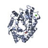
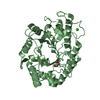
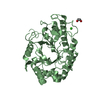
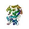
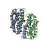
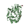
| Title | CATALYTIC CORE OF XYLANASE A E246C MUTANT | ||||||
 Components Components | XYLANASE A | ||||||
 Keywords Keywords | HYDROLASE / FAMILY F XYLANASE / FAMILY 10 OF GLYCOSYL-HYDROLASE | ||||||
| Function / homology |  Function and homology information Function and homology informationcellulose binding / endo-1,4-beta-xylanase / endo-1,4-beta-xylanase activity / xylan catabolic process Similarity search - Function | ||||||
| Biological species |  Cellvibrio japonicus (bacteria) Cellvibrio japonicus (bacteria) | ||||||
| Method |  X-RAY DIFFRACTION / Resolution: 2.5 Å X-RAY DIFFRACTION / Resolution: 2.5 Å | ||||||
 Authors Authors | Harris, G.W. / Jenkins, J.A. / Connerton, I. / Pickersgill, R.W. | ||||||
 Citation Citation | Journal: Structure / Year: 1994 Title: Structure of the catalytic core of the family F xylanase from Pseudomonas fluorescens and identification of the xylopentaose-binding sites. Authors: Harris, G.W. / Jenkins, J.A. / Connerton, I. / Cummings, N. / Lo Leggio, L. / Scott, M. / Hazlewood, G.P. / Laurie, J.I. / Gilbert, H.J. / Pickersgill, R.W. #1: Journal: FEBS Lett. / Year: 1995Title: Beta-Glucosidase, Beta-Galactosidase, Family a Cellulases, Family F Xylanases and Two Barley Glycanases Form a Superfamily of Enzymes with 8-Fold Beta-Alpha Architecture and with Two Conserved ...Title: Beta-Glucosidase, Beta-Galactosidase, Family a Cellulases, Family F Xylanases and Two Barley Glycanases Form a Superfamily of Enzymes with 8-Fold Beta-Alpha Architecture and with Two Conserved Glutamates Near the Carboxy-Terminal Ends of Beta-Strands Four and Seven Authors: Jenkins, J. / Lo Leggio, L. / Harris, G. / Pickersgill, R. #2: Journal: J.Mol.Biol. / Year: 1993Title: Crystallization and Preliminary X-Ray Analysis of the Catalytic Domain of Xylanase a from Pseudomonas Fluorescens Subspecies Cellulosa Authors: Pickersgill, R.W. / Jenkins, J.A. / Scott, M. / Connerton, I. / Hazlewood, G.P. / Gilbert, H.J. | ||||||
| History |
|
- Structure visualization
| Structure viewer | Molecule: MolmilJmol/JSmol |
|---|
- Downloads & links
-Download
| PDBx/mmCIF format | 1xys.cif.gz | 32.1 KB | Display | PDBx/mmCIF format |
|---|---|---|---|---|
| PDB format | pdb1xys.ent.gz | 17.6 KB | Display | PDB format |
| PDBx/mmJSON format | 1xys.json.gz | Tree view | PDBx/mmJSON format | |
| Others |  Other downloads Other downloads |
-Validation report
| Arichive directory | https://data.pdbj.org/pub/pdb/validation_reports/xy/1xysftp://data.pdbj.org/pub/pdb/validation_reports/xy/1xys | HTTPS FTP |
|---|
-Related structure data
| Similar structure data |
|---|


-Links
 PDBj
PDBj

- Assembly
| Deposited unit | 
| ||||||||
|---|---|---|---|---|---|---|---|---|---|
| 1 | 
| ||||||||
| 2 | 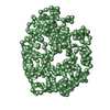
| ||||||||
| Unit cell |
| ||||||||
| Atom site foot note | 1: CIS PROLINE - PRO 221 | ||||||||
| Noncrystallographic symmetry (NCS) | NCS oper: (Code: given Matrix: (-0.9971, 0.0589, 0.0475), Vector: Details | MTRIX THE TRANSFORMATIONS PRESENTED ON MTRIX RECORDS BELOW DESCRIBE NON-CRYSTALLOGRAPHIC RELATIONSHIPS AMONG THE VARIOUS DOMAINS IN THIS ENTRY. APPLYING THE APPROPRIATE MTRIX TRANSFORMATION TO THE RESIDUES LISTED FIRST WILL YIELD APPROXIMATE COORDINATES FOR THE RESIDUES LISTED SECOND. APPLIED TO TRANSFORMED TO MTRIX RESIDUES RESIDUES RMSD M1 1 .. 345 1 .. 345 | |
-Components
| #1: Protein | Mass: 38434.508 Da / Num. of mol.: 2 Source method: isolated from a genetically manipulated source Source: (gene. exp.) Cellvibrio japonicus (bacteria) / Strain: CELLULOSA / Gene: TRUNCATED XYNA (CODONS 264-611) / Plasmid: PET3A / Gene (production host): TRUNCATED XYNA (CODONS 264-611) / Production host: #2: Chemical |   Mass: 40.078 Da / Num. of mol.: 2 / Source method: obtained synthetically / Formula: Ca Mass: 40.078 Da / Num. of mol.: 2 / Source method: obtained synthetically / Formula: CaCompound details | COMPND MOLECULE: XYLANASE A. CATALYTIC DOMAIN. | Nonpolymer details | CALCIUM 348 IS BOUND TO ASP 256, ASN 261, ASN 253, AND ASN 258. | |
|---|
-Experimental details
-Experiment
| Experiment | Method: X-RAY DIFFRACTION |
|---|
- Sample preparation
| Crystal | Density Matthews: 2.35 Å3/Da / Density % sol: 47.73 % | ||||||||||||||||||||||||||||||
|---|---|---|---|---|---|---|---|---|---|---|---|---|---|---|---|---|---|---|---|---|---|---|---|---|---|---|---|---|---|---|---|
| Crystal | *PLUS Density % sol: 47 % | ||||||||||||||||||||||||||||||
| Crystal grow | *PLUS Temperature: 18 ℃ / pH: 6.5 / Method: vapor diffusion, hanging drop | ||||||||||||||||||||||||||||||
| Components of the solutions | *PLUS
|
-Data collection
| Detector | Date: Dec 27, 1993 |
|---|---|
| Radiation | Monochromatic (M) / Laue (L): M / Scattering type: x-ray |
| Radiation wavelength | Relative weight: 1 |
| Reflection | Resolution: 2.5→35 Å / Num. obs: 25370 / % possible obs: 97.7 % / Observed criterion σ(I): 0 / Redundancy: 5.3 % / Rmerge(I) obs: 0.078 |
- Processing
| Software |
| |||||||||||||||||||||||||||||||||||||||||||||||||||||||||||||||
|---|---|---|---|---|---|---|---|---|---|---|---|---|---|---|---|---|---|---|---|---|---|---|---|---|---|---|---|---|---|---|---|---|---|---|---|---|---|---|---|---|---|---|---|---|---|---|---|---|---|---|---|---|---|---|---|---|---|---|---|---|---|---|---|---|
| Refinement | Resolution: 2.5→10 Å / σ(F): 0 /
| |||||||||||||||||||||||||||||||||||||||||||||||||||||||||||||||
| Displacement parameters | Biso mean: 14.4 Å2 | |||||||||||||||||||||||||||||||||||||||||||||||||||||||||||||||
| Refinement step | Cycle: LAST / Resolution: 2.5→10 Å
| |||||||||||||||||||||||||||||||||||||||||||||||||||||||||||||||
| Refine LS restraints |
|
