 Movie
Movie Controller

[English] 日本語
 Yorodumi
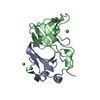
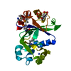
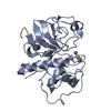
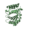
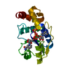
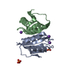
- PDB-1e44: ribonuclease domain of colicin E3 in complex with its immunity protein -
+ Open data

- Basic information
| Entry | Database: PDB / ID: 1.0E+44 | ||||||
|---|---|---|---|---|---|---|---|
| Title | ribonuclease domain of colicin E3 in complex with its immunity protein | ||||||
 Components Components |
| ||||||
 Keywords Keywords | RIBONUCLEASE / INHIBITION / PROTEIN-PROTEIN INTERACTIONS / RIBOSOME INACTIVATION / TOXIN | ||||||
| Function / homology |  Function and homology information Function and homology informationextrachromosomal circular DNA / bacteriocin immunity / hydrolase activity, acting on ester bonds / toxic substance binding / ribosome binding / Lyases; Phosphorus-oxygen lyases / endonuclease activity / killing of cells of another organism / transmembrane transporter binding / tRNA binding ...extrachromosomal circular DNA / bacteriocin immunity / hydrolase activity, acting on ester bonds / toxic substance binding / ribosome binding / Lyases; Phosphorus-oxygen lyases / endonuclease activity / killing of cells of another organism / transmembrane transporter binding / tRNA binding / lyase activity / defense response to bacterium / rRNA binding / extracellular region Similarity search - Function | ||||||
| Biological species |  | ||||||
| Method |  X-RAY DIFFRACTION / SYNCHROTRON / MAD / Resolution: 2.4 Å X-RAY DIFFRACTION / SYNCHROTRON / MAD / Resolution: 2.4 Å | ||||||
 Authors Authors | Carr, S. / Walker, D. / James, R. / Kleanthous, C. / Hemmings, A.M. | ||||||
 Citation Citation | Journal: Structure / Year: 2000 Title: Inhibition of a Ribosome Inactivating Ribonuclease: The Crystal Structure of the Cytotoxic Domain of Colicin E3 in Complex with its Immunity Protein Authors: Carr, S. / Walker, D. / James, R. / Kleanthous, C. / Hemmings, A.M. | ||||||
| History |
|
- Structure visualization
| Structure viewer | Molecule: MolmilJmol/JSmol |
|---|
- Downloads & links
-Download
| PDBx/mmCIF format | 1e44.cif.gz | 51.4 KB | Display | PDBx/mmCIF format |
|---|---|---|---|---|
| PDB format | pdb1e44.ent.gz | 37.5 KB | Display | PDB format |
| PDBx/mmJSON format | 1e44.json.gz | Tree view | PDBx/mmJSON format | |
| Others |  Other downloads Other downloads |
-Validation report
| Arichive directory | https://data.pdbj.org/pub/pdb/validation_reports/e4/1e44ftp://data.pdbj.org/pub/pdb/validation_reports/e4/1e44 | HTTPS FTP |
|---|
-Related structure data
| Similar structure data |
|---|


-Links
 PDBj
PDBj- Assembly
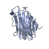
| Deposited unit | 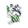
| ||||||||
|---|---|---|---|---|---|---|---|---|---|
| 1 |
| ||||||||
| Unit cell |
|
-Components
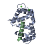
| #1: Protein | Mass: 9910.762 Da / Num. of mol.: 1 Source method: isolated from a genetically manipulated source Source: (gene. exp.) |
|---|---|
| #2: Protein | Mass: 10812.117 Da / Num. of mol.: 1 / Fragment: RIBONUCLEASE DOMAIN RESIDUES 456-551 Source method: isolated from a genetically manipulated source Source: (gene. exp.) Description: CO-EXPRESSED WITH A HIS-TAGGED IMMUNITY PROTEIN Cellular location (production host): CYTOPLASM / Production host: |
| #3: Chemical | ChemComp-EDO /   Mass: 62.068 Da / Num. of mol.: 1 / Source method: obtained synthetically / Formula: C2H6O2 Mass: 62.068 Da / Num. of mol.: 1 / Source method: obtained synthetically / Formula: C2H6O2 |
| #4: Water | ChemComp-HOH /  Mass: 18.015 Da / Num. of mol.: 171 / Source method: isolated from a natural source / Formula: H2O Mass: 18.015 Da / Num. of mol.: 171 / Source method: isolated from a natural source / Formula: H2O |
-Experimental details
-Experiment
| Experiment | Method: X-RAY DIFFRACTION / Number of used crystals: 1 |
|---|
- Sample preparation
| Crystal | Density Matthews: 4.39 Å3/Da / Density % sol: 72 % | ||||||||||||||||||||||||||||||
|---|---|---|---|---|---|---|---|---|---|---|---|---|---|---|---|---|---|---|---|---|---|---|---|---|---|---|---|---|---|---|---|
| Crystal grow | pH: 5.6 Details: 0.1 M NA CITRATE PH 5.6, 20% ISOPROPANOL, 10% PEG 4000 | ||||||||||||||||||||||||||||||
| Crystal grow | *PLUS Temperature: 277 K / pH: 7.5 / Method: vapor diffusion, hanging drop / Details: Carr, S., (1999) Acta Crystallogr., D56, 1630. | ||||||||||||||||||||||||||||||
| Components of the solutions | *PLUS
|
-Data collection
| Diffraction | Mean temperature: 100 K | ||||||||||||
|---|---|---|---|---|---|---|---|---|---|---|---|---|---|
| Diffraction source | Source: SYNCHROTRON / Site: ESRF  / Beamline: BM14 / Wavelength: 0.9, 0.97, 0.979 / Beamline: BM14 / Wavelength: 0.9, 0.97, 0.979 | ||||||||||||
| Detector | Detector: CCD | ||||||||||||
| Radiation | Protocol: MAD / Monochromatic (M) / Laue (L): M / Scattering type: x-ray | ||||||||||||
| Radiation wavelength |
| ||||||||||||
| Reflection | Resolution: 2.4→20 Å / Num. obs: 14892 / % possible obs: 98.7 % / Redundancy: 3 % / Rsym value: 0.039 / Net I/σ(I): 37.8 | ||||||||||||
| Reflection shell | Resolution: 2.4→2.45 Å / Redundancy: 2.7 % / Mean I/σ(I) obs: 2.8 / Rsym value: 0.237 / % possible all: 97.5 | ||||||||||||
| Reflection | *PLUS Lowest resolution: 20 Å / Rmerge(I) obs: 0.039 | ||||||||||||
| Reflection shell | *PLUS % possible obs: 97.5 % / Rmerge(I) obs: 0.237 |
- Processing
| Software |
| ||||||||||||||||||||||||||||||||||||||||||||||||||||||||||||
|---|---|---|---|---|---|---|---|---|---|---|---|---|---|---|---|---|---|---|---|---|---|---|---|---|---|---|---|---|---|---|---|---|---|---|---|---|---|---|---|---|---|---|---|---|---|---|---|---|---|---|---|---|---|---|---|---|---|---|---|---|---|
| Refinement | Method to determine structure: MAD / Resolution: 2.4→30 Å / Data cutoff high absF: 100000 / Cross valid method: THROUGHOUT / σ(F): 2
| ||||||||||||||||||||||||||||||||||||||||||||||||||||||||||||
| Refinement step | Cycle: LAST / Resolution: 2.4→30 Å
| ||||||||||||||||||||||||||||||||||||||||||||||||||||||||||||
| Refine LS restraints |
| ||||||||||||||||||||||||||||||||||||||||||||||||||||||||||||
| Software | *PLUS Name: CNS / Version: 0.5 / Classification: refinement | ||||||||||||||||||||||||||||||||||||||||||||||||||||||||||||
| Refinement | *PLUS Lowest resolution: 30 Å / σ(F): 2 | ||||||||||||||||||||||||||||||||||||||||||||||||||||||||||||
| Solvent computation | *PLUS | ||||||||||||||||||||||||||||||||||||||||||||||||||||||||||||
| Displacement parameters | *PLUS | ||||||||||||||||||||||||||||||||||||||||||||||||||||||||||||
| LS refinement shell | *PLUS Highest resolution: 2.4 Å / Lowest resolution: 2.45 Å / Rfactor Rfree: 0.229 / Rfactor obs: 0.194 |
