 Movie
Movie Controller


+ Open data

- Basic information
| Entry | Database: PDB / ID: 2w4d | ||||||
|---|---|---|---|---|---|---|---|
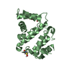
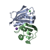
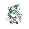
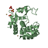
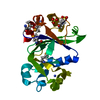
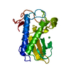
| Title | Acylphosphatase variant G91A from Pyrococcus horikoshii | ||||||
 Components Components | ACYLPHOSPHATASE | ||||||
 Keywords Keywords | HYDROLASE | ||||||
| Function / homology |  Function and homology information Function and homology information | ||||||
| Biological species |   PYROCOCCUS HORIKOSHII (archaea) PYROCOCCUS HORIKOSHII (archaea) | ||||||
| Method |  X-RAY DIFFRACTION / MOLECULAR REPLACEMENT / Resolution: 2.4 Å X-RAY DIFFRACTION / MOLECULAR REPLACEMENT / Resolution: 2.4 Å | ||||||
 Authors Authors | Lam, S.Y. / Wong, K.B. | ||||||
 Citation Citation | Journal: Plos Biol. / Year: 2011 Title: A Rigidifying Salt-Bridge Favors the Activity of Thermophilic Enzyme at High Temperatures at the Expense of Low-Temperature Activity. Authors: Lam, S.Y. / Yeung, R.C.Y. / Yu, T. / Sze, K. / Wong, K.B. #1: Journal: Acta Crystallogr.,Sect.D / Year: 2004 Title: Crystallization and Preliminary Crystallographic Analysis of an Acylphosphatase from the Hyperthermophilic Archaeon Pyrococcus Horikoshii Authors: Cheung, Y.Y. / Allen, M.D. / Bycroft, M. / Wong, K.B. #2: Journal: Biochemistry / Year: 2005Title: Crystal Structure of a Hyperthermophilic Archaeal Acylphosphatase from Pyrococcus Horikoshii--Structural Insights Into Enzymatic Catalysis, Thermostability, and Dimerization. Authors: Cheung, Y. / Lam, S.Y. / Chu, W. / Allen, M.D. / Bycroft, M. / Wong, K. | ||||||
| History |
|
- Structure visualization
| Structure viewer | Molecule: MolmilJmol/JSmol |
|---|
- Downloads & links
-Download
| PDBx/mmCIF format | 2w4d.cif.gz | 121.1 KB | Display | PDBx/mmCIF format |
|---|---|---|---|---|
| PDB format | pdb2w4d.ent.gz | 95.2 KB | Display | PDB format |
| PDBx/mmJSON format | 2w4d.json.gz | Tree view | PDBx/mmJSON format | |
| Others |  Other downloads Other downloads |
-Validation report
| Arichive directory | https://data.pdbj.org/pub/pdb/validation_reports/w4/2w4dftp://data.pdbj.org/pub/pdb/validation_reports/w4/2w4d | HTTPS FTP |
|---|
-Related structure data
| Related structure data |  2vh7C 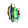 2w4cC 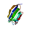 2w4pC 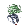 1w2iS S: Starting model for refinement C: citing same article ( |
|---|---|
| Similar structure data |


-Links
 PDBj
PDBj- Assembly
| Deposited unit | 
| ||||||||
|---|---|---|---|---|---|---|---|---|---|
| 1 | 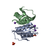
| ||||||||
| 2 | 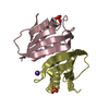
| ||||||||
| 3 | 
| ||||||||
| Unit cell |
|
-Components
| #1: Protein | Mass: 10159.682 Da / Num. of mol.: 6 / Fragment: RESIDUES 2-91 / Mutation: YES Source method: isolated from a genetically manipulated source Source: (gene. exp.) PYROCOCCUS HORIKOSHII (archaea) / Strain: JCM9974 / Plasmid: PET3A / Production host:  #2: Chemical | ChemComp-K /   Mass: 39.098 Da / Num. of mol.: 6 / Source method: obtained synthetically / Formula: K Mass: 39.098 Da / Num. of mol.: 6 / Source method: obtained synthetically / Formula: K#3: Chemical | ChemComp-PO4 /   Mass: 94.971 Da / Num. of mol.: 7 / Source method: obtained synthetically / Formula: PO4 Mass: 94.971 Da / Num. of mol.: 7 / Source method: obtained synthetically / Formula: PO4#4: Water | ChemComp-HOH / |  Mass: 18.015 Da / Num. of mol.: 125 / Source method: isolated from a natural source / Formula: H2O Mass: 18.015 Da / Num. of mol.: 125 / Source method: isolated from a natural source / Formula: H2OCompound details | ENGINEERED RESIDUE IN CHAIN A, GLY 91 TO ALA ENGINEERED RESIDUE IN CHAIN B, GLY 91 TO ALA ...ENGINEERED | |
|---|
-Experimental details
-Experiment
| Experiment | Method: X-RAY DIFFRACTION / Number of used crystals: 1 |
|---|
- Sample preparation
| Crystal | Density Matthews: 2.5 Å3/Da / Density % sol: 50 % / Description: NONE |
|---|---|
| Crystal grow | pH: 8.5 Details: 7.9MG/ML PROTEIN, 0.6M NA/K GRID SCREEN, PH8.5, CRYOPROTECTANT: 25% GLYCEROL |
-Data collection
| Diffraction | Mean temperature: 289 K |
|---|---|
| Diffraction source | Source: ROTATING ANODE / Type: RIGAKU MICROMAX-007 / Wavelength: 1.5 |
| Detector | Type: RIGAKU IMAGE PLATE / Detector: IMAGE PLATE / Date: Jul 20, 2005 / Details: MIRRORS |
| Radiation | Protocol: SINGLE WAVELENGTH / Monochromatic (M) / Laue (L): M / Scattering type: x-ray |
| Radiation wavelength | Wavelength: 1.5 Å / Relative weight: 1 |
| Reflection | Resolution: 2.4→44.18 Å / Num. obs: 24753 / % possible obs: 98.9 % / Observed criterion σ(I): 2 / Redundancy: 2.92 % / Biso Wilson estimate: 27.7 Å2 / Rmerge(I) obs: 0.07 / Net I/σ(I): 10.7 |
| Reflection shell | Resolution: 2.4→2.49 Å / Redundancy: 2.74 % / Rmerge(I) obs: 0.2 / Mean I/σ(I) obs: 4.2 / % possible all: 97.5 |
- Processing
| Software |
| ||||||||||||||||||||||||||||||||||||||||||||||||||||||||||||
|---|---|---|---|---|---|---|---|---|---|---|---|---|---|---|---|---|---|---|---|---|---|---|---|---|---|---|---|---|---|---|---|---|---|---|---|---|---|---|---|---|---|---|---|---|---|---|---|---|---|---|---|---|---|---|---|---|---|---|---|---|---|
| Refinement | Method to determine structure: MOLECULAR REPLACEMENT Starting model: PDB ENTRY 1W2I Resolution: 2.4→23.44 Å / Rfactor Rfree error: 0.007 / Data cutoff high absF: 1417720.6 / Isotropic thermal model: RESTRAINED / Cross valid method: THROUGHOUT / σ(F): 0
| ||||||||||||||||||||||||||||||||||||||||||||||||||||||||||||
| Solvent computation | Solvent model: FLAT MODEL / Bsol: 39.35 Å2 / ksol: 0.37 e/Å3 | ||||||||||||||||||||||||||||||||||||||||||||||||||||||||||||
| Displacement parameters | Biso mean: 32.7 Å2
| ||||||||||||||||||||||||||||||||||||||||||||||||||||||||||||
| Refine analyze |
| ||||||||||||||||||||||||||||||||||||||||||||||||||||||||||||
| Refinement step | Cycle: LAST / Resolution: 2.4→23.44 Å
| ||||||||||||||||||||||||||||||||||||||||||||||||||||||||||||
| Refine LS restraints |
| ||||||||||||||||||||||||||||||||||||||||||||||||||||||||||||
| Refine LS restraints NCS | NCS model details: CONSTR | ||||||||||||||||||||||||||||||||||||||||||||||||||||||||||||
| LS refinement shell | Resolution: 2.4→2.55 Å / Rfactor Rfree error: 0.024 / Total num. of bins used: 6
| ||||||||||||||||||||||||||||||||||||||||||||||||||||||||||||
| Xplor file |
|
