 Movie
Movie Controller


+ Open data

- Basic information
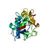
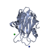
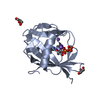
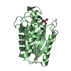
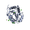
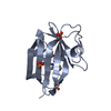
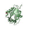
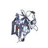
| Entry | Database: PDB / ID: 1dy0 | ||||||
|---|---|---|---|---|---|---|---|
| Title | Murine endostatin, crystal form II | ||||||
 Components Components | COLLAGEN ALPHA1(XVIII) CHAIN | ||||||
 Keywords Keywords | ANGIOGENESIS INHIBITOR | ||||||
| Function / homology |  Function and homology information Function and homology informationActivation of Matrix Metalloproteinases / Collagen biosynthesis and modifying enzymes / Collagen chain trimerization / Collagen degradation / Assembly of collagen fibrils and other multimeric structures / extracellular matrix structural constituent conferring tensile strength / Integrin cell surface interactions / endothelial cell morphogenesis / collagen trimer / basement membrane ...Activation of Matrix Metalloproteinases / Collagen biosynthesis and modifying enzymes / Collagen chain trimerization / Collagen degradation / Assembly of collagen fibrils and other multimeric structures / extracellular matrix structural constituent conferring tensile strength / Integrin cell surface interactions / endothelial cell morphogenesis / collagen trimer / basement membrane / positive regulation of endothelial cell apoptotic process / extracellular matrix organization / skeletal system development / extracellular matrix / angiogenesis / cell adhesion / positive regulation of cell migration / positive regulation of cell population proliferation / : / metal ion binding / identical protein binding Similarity search - Function | ||||||
| Biological species |  | ||||||
| Method |  X-RAY DIFFRACTION / MOLECULAR REPLACEMENT / Resolution: 2.2 Å X-RAY DIFFRACTION / MOLECULAR REPLACEMENT / Resolution: 2.2 Å | ||||||
 Authors Authors | Hohenester, E. / Sasaki, T. / Timpl, R. | ||||||
 Citation Citation | Journal: J.Mol.Biol. / Year: 2000 Title: Variable Zinc Coordination in Endostatin Authors: Hohenester, E. / Sasaki, T. / Mann, K. / Timpl, R. #1: Journal: Embo J. / Year: 1998Title: Crystal Structure of the Angiogenesis Inhibitor Endostatin at 1.5 Angstrom Resolution Authors: Hohenester, E. / Sasaki, T. / Olsen, B.R. / Timpl, R. | ||||||
| History |
|
- Structure visualization
| Structure viewer | Molecule: MolmilJmol/JSmol |
|---|
- Downloads & links
-Download
| PDBx/mmCIF format | 1dy0.cif.gz | 49.2 KB | Display | PDBx/mmCIF format |
|---|---|---|---|---|
| PDB format | pdb1dy0.ent.gz | 34.3 KB | Display | PDB format |
| PDBx/mmJSON format | 1dy0.json.gz | Tree view | PDBx/mmJSON format | |
| Others |  Other downloads Other downloads |
-Validation report
| Arichive directory | https://data.pdbj.org/pub/pdb/validation_reports/dy/1dy0ftp://data.pdbj.org/pub/pdb/validation_reports/dy/1dy0 | HTTPS FTP |
|---|
-Related structure data
| Related structure data |  1dy1C  1koeS S: Starting model for refinement C: citing same article ( |
|---|---|
| Similar structure data |


-Links
 PDBj
PDBj


- Assembly
| Deposited unit | 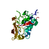
| ||||||||
|---|---|---|---|---|---|---|---|---|---|
| 1 |
| ||||||||
| Unit cell |
| ||||||||
| Details | BIOLOGICAL_UNIT: MONOMER |
-Components
| #1: Protein | Mass: 20753.387 Da / Num. of mol.: 1 / Fragment: ENDOSTATIN DOMAIN / Mutation: YES Source method: isolated from a genetically manipulated source Source: (gene. exp.)  HOMO SAPIENS (human) / References: UniProt: P39061 HOMO SAPIENS (human) / References: UniProt: P39061 | ||||||
|---|---|---|---|---|---|---|---|
| #2: Chemical |   Mass: 65.409 Da / Num. of mol.: 2 / Source method: obtained synthetically / Formula: Zn Mass: 65.409 Da / Num. of mol.: 2 / Source method: obtained synthetically / Formula: Zn#3: Water | ChemComp-HOH / |  Mass: 18.015 Da / Num. of mol.: 44 / Source method: isolated from a natural source / Formula: H2O Mass: 18.015 Da / Num. of mol.: 44 / Source method: isolated from a natural source / Formula: H2OHas protein modification | Y | Sequence details | N-TERMINAL APLA MUTATION | |
-Experimental details
-Experiment
| Experiment | Method: X-RAY DIFFRACTION / Number of used crystals: 1 |
|---|
- Sample preparation
| Crystal | Density Matthews: 2.09 Å3/Da / Density % sol: 41.22 % | ||||||||||||||||||||||||||||||||||||||||||
|---|---|---|---|---|---|---|---|---|---|---|---|---|---|---|---|---|---|---|---|---|---|---|---|---|---|---|---|---|---|---|---|---|---|---|---|---|---|---|---|---|---|---|---|
| Crystal grow | pH: 8.5 / Details: pH 8.50 | ||||||||||||||||||||||||||||||||||||||||||
| Crystal grow | *PLUS pH: 8 / Method: vapor diffusion, hanging drop | ||||||||||||||||||||||||||||||||||||||||||
| Components of the solutions | *PLUS
|
-Data collection
| Diffraction | Mean temperature: 293 K |
|---|---|
| Diffraction source | Source: ROTATING ANODE / Type: ELLIOTT GX-21 / Wavelength: 1.5418 / Wavelength: 1.5418 Å |
| Detector | Type: MARRESEARCH / Detector: IMAGE PLATE / Date: Mar 15, 1998 |
| Radiation | Monochromator: GRAPHITE(002) / Protocol: SINGLE WAVELENGTH / Monochromatic (M) / Laue (L): M / Scattering type: x-ray |
| Radiation wavelength | Wavelength: 1.5418 Å / Relative weight: 1 |
| Reflection | Resolution: 2.2→15 Å / Num. obs: 9450 / % possible obs: 99.8 % / Redundancy: 8.1 % / Rmerge(I) obs: 0.079 |
| Reflection shell | Resolution: 2.2→2.32 Å / Rmerge(I) obs: 0.197 / % possible all: 99.9 |
| Reflection | *PLUS Num. measured all: 76689 |
| Reflection shell | *PLUS % possible obs: 99.8 % |
- Processing
| Software |
| ||||||||||||||||||||||||||||||||||||||||||||||||||||||||||||
|---|---|---|---|---|---|---|---|---|---|---|---|---|---|---|---|---|---|---|---|---|---|---|---|---|---|---|---|---|---|---|---|---|---|---|---|---|---|---|---|---|---|---|---|---|---|---|---|---|---|---|---|---|---|---|---|---|---|---|---|---|---|
| Refinement | Method to determine structure: MOLECULAR REPLACEMENT Starting model: PDB ENTRY 1KOE Resolution: 2.2→8 Å / Isotropic thermal model: INDIVIDUAL RESTRAINED / Cross valid method: THROUGHOUT / σ(F): 0
| ||||||||||||||||||||||||||||||||||||||||||||||||||||||||||||
| Refinement step | Cycle: LAST / Resolution: 2.2→8 Å
| ||||||||||||||||||||||||||||||||||||||||||||||||||||||||||||
| Refine LS restraints |
|
