 Movie
Movie Controller


+ Open data

- Basic information
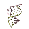
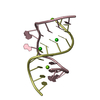
| Entry | Database: PDB / ID: 1duq | ||||||
|---|---|---|---|---|---|---|---|
| Title | CRYSTAL STRUCTURE OF THE REV BINDING ELEMENT OF HIV-1 | ||||||
 Components Components | (THE REV BINDING ELEMENT) x 2 | ||||||
 Keywords Keywords | RNA / RRE / HIV-1 / REV BINDING DOMAIN | ||||||
| Function / homology | RNA / RNA (> 10) Function and homology information Function and homology information | ||||||
| Method |  X-RAY DIFFRACTION / SYNCHROTRON / Resolution: 2.1 Å X-RAY DIFFRACTION / SYNCHROTRON / Resolution: 2.1 Å | ||||||
 Authors Authors | Hung, L.-W. / Holbrook, E.L. / Holbrook, S.R. | ||||||
 Citation Citation | Journal: Proc.Natl.Acad.Sci.USA / Year: 2000 Title: The crystal structure of the Rev binding element of HIV-1 reveals novel base pairing and conformational variability. Authors: Hung, L.W. / Holbrook, E.L. / Holbrook, S.R. | ||||||
| History |
|
- Structure visualization
| Structure viewer | Molecule: MolmilJmol/JSmol |
|---|
- Downloads & links
-Download
| PDBx/mmCIF format | 1duq.cif.gz | 70.8 KB | Display | PDBx/mmCIF format |
|---|---|---|---|---|
| PDB format | pdb1duq.ent.gz | 51.1 KB | Display | PDB format |
| PDBx/mmJSON format | 1duq.json.gz | Tree view | PDBx/mmJSON format | |
| Others |  Other downloads Other downloads |
-Validation report
| Arichive directory | https://data.pdbj.org/pub/pdb/validation_reports/du/1duqftp://data.pdbj.org/pub/pdb/validation_reports/du/1duq | HTTPS FTP |
|---|
-Related structure data
| Similar structure data |
|---|


-Links
 PDBj
PDBj


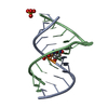
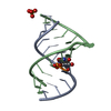
- Assembly
| Deposited unit | 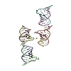
| ||||||||||
|---|---|---|---|---|---|---|---|---|---|---|---|
| 1 | 
| ||||||||||
| 2 | 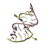
| ||||||||||
| 3 | 
| ||||||||||
| 4 | 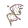
| ||||||||||
| Unit cell |
|
-Components
| #1: RNA chain | Mass: 3922.400 Da / Num. of mol.: 4 / Fragment: SHORT CHAIN / Source method: obtained synthetically / Details: SEQUENCE FROM HIV-1. SYNTHESIZED BY OLIGOS, ETC. #2: RNA chain | Mass: 4461.723 Da / Num. of mol.: 4 / Fragment: LONG CHAIN / Source method: obtained synthetically #3: Chemical | ChemComp-NA /   Mass: 22.990 Da / Num. of mol.: 10 / Source method: obtained synthetically / Formula: Na Mass: 22.990 Da / Num. of mol.: 10 / Source method: obtained synthetically / Formula: Na#4: Water | ChemComp-HOH / |  Mass: 18.015 Da / Num. of mol.: 83 / Source method: isolated from a natural source / Formula: H2O Mass: 18.015 Da / Num. of mol.: 83 / Source method: isolated from a natural source / Formula: H2O |
|---|
-Experimental details
-Experiment
| Experiment | Method: X-RAY DIFFRACTION / Number of used crystals: 1 |
|---|
- Sample preparation
| Crystal | Density Matthews: 2.2 Å3/Da / Density % sol: 44.19 % | ||||||||||||||||||||||||||||||||||||
|---|---|---|---|---|---|---|---|---|---|---|---|---|---|---|---|---|---|---|---|---|---|---|---|---|---|---|---|---|---|---|---|---|---|---|---|---|---|
| Crystal grow | Temperature: 294 K / Method: vapor diffusion, hanging drop / pH: 6.5 Details: 0.1M SODIUM CACODYLATE, PH 6.5, 0.2 M NACL, 35% MPD, VAPOR DIFFUSION, HANGING DROP, temperature 294K | ||||||||||||||||||||||||||||||||||||
| Components of the solutions |
| ||||||||||||||||||||||||||||||||||||
| Crystal grow | *PLUS | ||||||||||||||||||||||||||||||||||||
| Components of the solutions | *PLUS
|
-Data collection
| Diffraction | Mean temperature: 100 K |
|---|---|
| Diffraction source | Source: SYNCHROTRON / Site: ALS  / Beamline: 5.0.2 / Wavelength: 1.1 / Beamline: 5.0.2 / Wavelength: 1.1 |
| Detector | Type: ADSC QUANTUM 4 / Detector: CCD / Date: Aug 26, 1999 |
| Radiation | Protocol: SINGLE WAVELENGTH / Monochromatic (M) / Laue (L): M / Scattering type: x-ray |
| Radiation wavelength | Wavelength: 1.1 Å / Relative weight: 1 |
| Reflection | Resolution: 2.1→20 Å / Num. all: 16703 / Num. obs: 15998 / % possible obs: 94.7 % / Observed criterion σ(F): 2 / Observed criterion σ(I): 1 / Redundancy: 1.8 % / Biso Wilson estimate: 25.6 Å2 / Rmerge(I) obs: 0.38 / Net I/σ(I): 8 |
| Reflection shell | Resolution: 2.1→2.17 Å / Redundancy: 1.5 % / Rmerge(I) obs: 0.248 / % possible all: 83.2 |
| Reflection shell | *PLUS % possible obs: 83 % |
- Processing
| Software |
| ||||||||||||||||||||||||||||||||||||||||||||||||||||||||||||
|---|---|---|---|---|---|---|---|---|---|---|---|---|---|---|---|---|---|---|---|---|---|---|---|---|---|---|---|---|---|---|---|---|---|---|---|---|---|---|---|---|---|---|---|---|---|---|---|---|---|---|---|---|---|---|---|---|---|---|---|---|---|
| Refinement | Resolution: 2.1→19.77 Å / Rfactor Rfree error: 0.007 / Data cutoff high absF: 659115.35 / Data cutoff low absF: 0 / Isotropic thermal model: RESTRAINED / Cross valid method: THROUGHOUT / σ(F): 2 / σ(I): 1 / Stereochemistry target values: ENGH & HUBER
| ||||||||||||||||||||||||||||||||||||||||||||||||||||||||||||
| Solvent computation | Solvent model: FLAT MODEL / Bsol: 60.11 Å2 / ksol: 0.399 e/Å3 | ||||||||||||||||||||||||||||||||||||||||||||||||||||||||||||
| Displacement parameters | Biso mean: 41.1 Å2
| ||||||||||||||||||||||||||||||||||||||||||||||||||||||||||||
| Refine analyze |
| ||||||||||||||||||||||||||||||||||||||||||||||||||||||||||||
| Refinement step | Cycle: LAST / Resolution: 2.1→19.77 Å
| ||||||||||||||||||||||||||||||||||||||||||||||||||||||||||||
| Refine LS restraints |
| ||||||||||||||||||||||||||||||||||||||||||||||||||||||||||||
| LS refinement shell | Resolution: 2.1→2.23 Å / Rfactor Rfree error: 0.029 / Total num. of bins used: 6
| ||||||||||||||||||||||||||||||||||||||||||||||||||||||||||||
| Xplor file |
| ||||||||||||||||||||||||||||||||||||||||||||||||||||||||||||
| Software | *PLUS Name: CNS / Version: 0.9 / Classification: refinement | ||||||||||||||||||||||||||||||||||||||||||||||||||||||||||||
| Refinement | *PLUS σ(F): 2 / % reflection Rfree: 10.1 % | ||||||||||||||||||||||||||||||||||||||||||||||||||||||||||||
| Solvent computation | *PLUS | ||||||||||||||||||||||||||||||||||||||||||||||||||||||||||||
| Displacement parameters | *PLUS Biso mean: 41.1 Å2 | ||||||||||||||||||||||||||||||||||||||||||||||||||||||||||||
| Refine LS restraints | *PLUS
| ||||||||||||||||||||||||||||||||||||||||||||||||||||||||||||
| LS refinement shell | *PLUS Rfactor Rfree: 0.385 / % reflection Rfree: 10.1 % / Rfactor Rwork: 0.301 |
