 Movie
Movie Controller


+ Open data

- Basic information
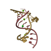
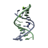
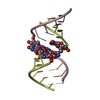
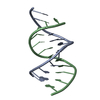
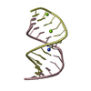
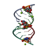
| Entry | Database: PDB / ID: 1nuj | ||||||
|---|---|---|---|---|---|---|---|
| Title | THE LEADZYME STRUCTURE BOUND TO MG(H20)6(II) AT 1.8 A RESOLUTION | ||||||
 Components Components |
| ||||||
 Keywords Keywords | RNA / RIBOZYME / LEADZYME / LEAD-DEPENDENT CLEAVAGE / MG(H20)62+ / BULGED NUCLEOTIDES / HYDRATED MAGNESIUM / PSEUDOHELICAL PACKING / STICKY ENDS / ALTERNATE CONFORMATION / HOMOPURINE BASE PAIRS | ||||||
| Function / homology | RNA / RNA (> 10) Function and homology information Function and homology information | ||||||
| Method |  X-RAY DIFFRACTION / SYNCHROTRON / EXPAND TO P6(1) / Resolution: 1.8 Å X-RAY DIFFRACTION / SYNCHROTRON / EXPAND TO P6(1) / Resolution: 1.8 Å | ||||||
 Authors Authors | Wedekind, J.E. / Mckay, D.B. | ||||||
 Citation Citation | Journal: BIOCHEMISTRY / Year: 2003 Title: Crystal structure of the leadzyme at 1.8 A resolution: metal ion binding and the implications for catalytic mechanism and allo site ion regulation. Authors: Wedekind, J.E. / McKay, D.B. #1: Journal: Nat.Struct.Biol. / Year: 1999Title: CRYSTAL STRUCTURE OF A LEAD DEPENDENT RIBOZYME REVEALING METAL BINDING SITES RELEVANT TO CATALYSIS Authors: Wedekind, J.E. / McKay, D.B. | ||||||
| History |
|
- Structure visualization
| Structure viewer | Molecule: MolmilJmol/JSmol |
|---|
- Downloads & links
-Download
| PDBx/mmCIF format | 1nuj.cif.gz | 75 KB | Display | PDBx/mmCIF format |
|---|---|---|---|---|
| PDB format | pdb1nuj.ent.gz | 54.8 KB | Display | PDB format |
| PDBx/mmJSON format | 1nuj.json.gz | Tree view | PDBx/mmJSON format | |
| Others |  Other downloads Other downloads |
-Validation report
| Arichive directory | https://data.pdbj.org/pub/pdb/validation_reports/nu/1nujftp://data.pdbj.org/pub/pdb/validation_reports/nu/1nuj | HTTPS FTP |
|---|
-Related structure data
| Related structure data | 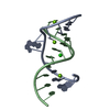 1nuvC C: citing same article ( |
|---|---|
| Similar structure data | |
| Other databases |
|


-Links
 PDBj
PDBj


- Assembly
| Deposited unit | 
| ||||||||
|---|---|---|---|---|---|---|---|---|---|
| 1 | 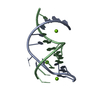
| ||||||||
| 2 | 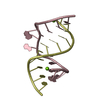
| ||||||||
| 3 | 
| ||||||||
| 4 | 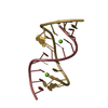
| ||||||||
| Unit cell |
|
-Components
| #1: RNA chain | Mass: 4194.598 Da / Num. of mol.: 4 / Source method: obtained synthetically Details: This ribozyme was made by in vitro selection, reported by Pan & Uhlenbeck. #2: RNA chain | Mass: 3538.154 Da / Num. of mol.: 4 / Source method: obtained synthetically Details: This ribozyme was made by in vitro selection, reported by Pan & Uhlenbeck. #3: Chemical | ChemComp-MG /   Mass: 24.305 Da / Num. of mol.: 10 / Source method: obtained synthetically / Formula: Mg Mass: 24.305 Da / Num. of mol.: 10 / Source method: obtained synthetically / Formula: Mg#4: Water | ChemComp-HOH / |  Mass: 18.015 Da / Num. of mol.: 416 / Source method: isolated from a natural source / Formula: H2O Mass: 18.015 Da / Num. of mol.: 416 / Source method: isolated from a natural source / Formula: H2O |
|---|
-Experimental details
-Experiment
| Experiment | Method: X-RAY DIFFRACTION / Number of used crystals: 1 |
|---|
- Sample preparation
| Crystal | Density Matthews: 1.98 Å3/Da / Density % sol: 37.52 % | |||||||||||||||||||||||||||||||||||
|---|---|---|---|---|---|---|---|---|---|---|---|---|---|---|---|---|---|---|---|---|---|---|---|---|---|---|---|---|---|---|---|---|---|---|---|---|
| Crystal grow | Temperature: 293 K / Method: vapor diffusion, hanging drop / pH: 6 Details: MPD, Mg-Acetate, cacodylate, pH 6.0, VAPOR DIFFUSION, HANGING DROP, temperature 20K | |||||||||||||||||||||||||||||||||||
| Components of the solutions |
| |||||||||||||||||||||||||||||||||||
| Crystal grow | *PLUS Temperature: 20 ℃ / Method: vapor diffusion, hanging drop | |||||||||||||||||||||||||||||||||||
| Components of the solutions | *PLUS
|
-Data collection
| Diffraction | Mean temperature: 100 K |
|---|---|
| Diffraction source | Source: SYNCHROTRON / Site: SSRL  / Beamline: BL9-1 / Wavelength: 0.98 Å / Beamline: BL9-1 / Wavelength: 0.98 Å |
| Detector | Type: MAR scanner 345 mm plate / Detector: IMAGE PLATE / Date: Mar 4, 1999 Details: 16 pole wiggler, Flat mirror, Si(311) bent mono with horizontal focus |
| Radiation | Monochromator: si(311) / Protocol: SINGLE WAVELENGTH / Monochromatic (M) / Laue (L): M / Scattering type: x-ray |
| Radiation wavelength | Wavelength: 0.98 Å / Relative weight: 1 |
| Reflection | Resolution: 1.8→34 Å / Num. obs: 25779 / % possible obs: 95.2 % / Observed criterion σ(I): -3 / Redundancy: 3.1 % / Biso Wilson estimate: 26.8 Å2 / Rsym value: 0.04 / Net I/σ(I): 21.3 |
| Reflection shell | Resolution: 1.8→1.83 Å / Redundancy: 2.1 % / Mean I/σ(I) obs: 4.6 / Rsym value: 0.272 / % possible all: 87.4 |
| Reflection | *PLUS Lowest resolution: 34 Å / Num. obs: 25179 / Num. measured all: 286623 / Rmerge(I) obs: 0.04 |
| Reflection shell | *PLUS Rmerge(I) obs: 0.272 |
- Processing
| Software |
| |||||||||||||||||||||||||
|---|---|---|---|---|---|---|---|---|---|---|---|---|---|---|---|---|---|---|---|---|---|---|---|---|---|---|
| Refinement | Method to determine structure: EXPAND TO P6(1) Starting model: NDB ENTRY UR0001 Resolution: 1.8→29.31 Å / Rfactor Rfree error: 0.007 / Isotropic thermal model: RESTRAINED / Cross valid method: THROUGHOUT / σ(F): 0 Stereochemistry target values: G. Parkinson, J. Vojtechovsky, L. Clowney, A.T. Brunger, H.M. Berman Details: RESOLUTION-DEPENDENT WEIGHTING SCHEME
| |||||||||||||||||||||||||
| Solvent computation | Solvent model: FLAT MODEL / Bsol: 42.4334 Å2 / ksol: 0.287285 e/Å3 | |||||||||||||||||||||||||
| Displacement parameters | Biso mean: 30.2 Å2
| |||||||||||||||||||||||||
| Refine analyze | Luzzati coordinate error free: 0.37 Å / Luzzati sigma a free: 0.38 Å | |||||||||||||||||||||||||
| Refinement step | Cycle: LAST / Resolution: 1.8→29.31 Å
| |||||||||||||||||||||||||
| Refine LS restraints |
| |||||||||||||||||||||||||
| LS refinement shell | Resolution: 1.8→1.86 Å / Rfactor Rfree error: 0.034 / Total num. of bins used: 10
| |||||||||||||||||||||||||
| Refinement | *PLUS Highest resolution: 1.8 Å / Lowest resolution: 34 Å / % reflection Rfree: 8 % | |||||||||||||||||||||||||
| Solvent computation | *PLUS | |||||||||||||||||||||||||
| Displacement parameters | *PLUS | |||||||||||||||||||||||||
| Refine LS restraints | *PLUS
|
