 Movie
Movie Controller

[English] 日本語
 Yorodumi
- PDB-3cgq: X-ray structure of a pseudouridine-containing yeast spliceosomal ... -
+ Open data

- Basic information
| Entry | Database: PDB / ID: 3cgq | ||||||
|---|---|---|---|---|---|---|---|
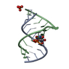
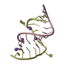
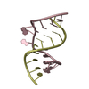
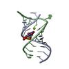
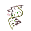
| Title | X-ray structure of a pseudouridine-containing yeast spliceosomal U2 snRNA-intron branch site duplex | ||||||
 Components Components |
| ||||||
 Keywords Keywords | RNA / RNA double helix / Branchpoint sequence / BPS / U2 snRNA / pseudouridine / pre-mRNA splicing | ||||||
| Function / homology | RNA / RNA (> 10) Function and homology information Function and homology information | ||||||
| Method |  X-RAY DIFFRACTION / MOLECULAR REPLACEMENT / Resolution: 2.55 Å X-RAY DIFFRACTION / MOLECULAR REPLACEMENT / Resolution: 2.55 Å | ||||||
 Authors Authors | Lin, Y. / Kielkopf, C.L. | ||||||
 Citation Citation | Journal: Biochemistry / Year: 2008 Title: X-ray structures of U2 snRNA-branchpoint duplexes containing conserved pseudouridines. Authors: Lin, Y. / Kielkopf, C.L. | ||||||
| History |
|
- Structure visualization
| Structure viewer | Molecule: MolmilJmol/JSmol |
|---|
- Downloads & links
-Download
| PDBx/mmCIF format | 3cgq.cif.gz | 24.5 KB | Display | PDBx/mmCIF format |
|---|---|---|---|---|
| PDB format | pdb3cgq.ent.gz | 15.7 KB | Display | PDB format |
| PDBx/mmJSON format | 3cgq.json.gz | Tree view | PDBx/mmJSON format | |
| Others |  Other downloads Other downloads |
-Validation report
| Arichive directory | https://data.pdbj.org/pub/pdb/validation_reports/cg/3cgqftp://data.pdbj.org/pub/pdb/validation_reports/cg/3cgq | HTTPS FTP |
|---|
-Related structure data
| Related structure data |  3cgpC  3cgrC  3cgsC 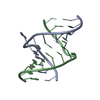 1i9xS 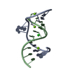 1nuvS C: citing same article ( S: Starting model for refinement |
|---|---|
| Similar structure data |


-Links
 PDBj
PDBj


- Assembly
| Deposited unit | 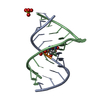
| ||||||||
|---|---|---|---|---|---|---|---|---|---|
| 1 |
| ||||||||
| Unit cell |
|
-Components
| #1: RNA chain | Mass: 3867.360 Da / Num. of mol.: 1 / Source method: obtained synthetically Details: Central nucleotides of this sequence are highly conserved in yeast U2 snRNA |
|---|---|
| #2: RNA chain | Mass: 4116.518 Da / Num. of mol.: 1 / Source method: obtained synthetically Details: Central nucleotides of this sequence are highly conserved in yeast pre-mRNAs |
| #3: Chemical | ChemComp-SO4 /   Mass: 96.063 Da / Num. of mol.: 1 / Source method: obtained synthetically / Formula: SO4 Mass: 96.063 Da / Num. of mol.: 1 / Source method: obtained synthetically / Formula: SO4 |
| #4: Water | ChemComp-HOH /  Mass: 18.015 Da / Num. of mol.: 49 / Source method: isolated from a natural source / Formula: H2O Mass: 18.015 Da / Num. of mol.: 49 / Source method: isolated from a natural source / Formula: H2O |
-Experimental details
-Experiment
| Experiment | Method: X-RAY DIFFRACTION / Number of used crystals: 1 |
|---|
- Sample preparation
| Crystal | Density Matthews: 2.9 Å3/Da / Density % sol: 57.56 % | ||||||||||||
|---|---|---|---|---|---|---|---|---|---|---|---|---|---|
| Crystal grow | Temperature: 298 K / Method: vapor diffusion, hanging drop / pH: 6.5 Details: 1M ammonium sulfate, pH 6.5, VAPOR DIFFUSION, HANGING DROP, temperature 298.0K | ||||||||||||
| Components of the solutions |
|
-Data collection
| Diffraction | Mean temperature: 100 K |
|---|---|
| Diffraction source | Source: ROTATING ANODE / Type: BRUKER AXS MICROSTAR / Wavelength: 1.54 Å |
| Detector | Detector: CCD / Date: Dec 21, 2004 |
| Radiation | Protocol: SINGLE WAVELENGTH / Monochromatic (M) / Laue (L): M / Scattering type: x-ray |
| Radiation wavelength | Wavelength: 1.54 Å / Relative weight: 1 |
| Reflection | Resolution: 2.5→20 Å / Num. all: 3054 / Num. obs: 3027 / % possible obs: 99.5 % / Observed criterion σ(F): 0 / Observed criterion σ(I): 0 / Redundancy: 6.8 % / Rsym value: 0.053 / Net I/σ(I): 44.1 |
| Reflection shell | Resolution: 2.55→2.64 Å / Redundancy: 6 % / Mean I/σ(I) obs: 16.5 / Rsym value: 0.108 / % possible all: 100 |
- Processing
| Software |
| |||||||||||||||||||||||||
|---|---|---|---|---|---|---|---|---|---|---|---|---|---|---|---|---|---|---|---|---|---|---|---|---|---|---|
| Refinement | Method to determine structure: MOLECULAR REPLACEMENT Starting model: Combination of PDB entry 1I9X and 1NUV Resolution: 2.55→20 Å / Cross valid method: THROUGHOUT / σ(F): 0 / σ(I): 0 / Stereochemistry target values: Engh & Huber
| |||||||||||||||||||||||||
| Displacement parameters | Biso mean: 15.1 Å2 | |||||||||||||||||||||||||
| Refine analyze | Luzzati coordinate error obs: 0.3 Å / Luzzati d res low obs: 5 Å / Luzzati sigma a obs: 0.33 Å | |||||||||||||||||||||||||
| Refinement step | Cycle: LAST / Resolution: 2.55→20 Å
| |||||||||||||||||||||||||
| Refine LS restraints |
|
