 Movie
Movie Controller

[English] 日本語
 Yorodumi
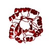
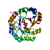
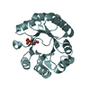
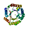
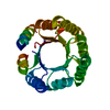
- PDB-1dl3: CRYSTAL STRUCTURE OF MUTUALLY GENERATED MONOMERS OF DIMERIC PHOSP... -
+ Open data

- Basic information
| Entry | Database: PDB / ID: 1dl3 | ||||||
|---|---|---|---|---|---|---|---|
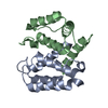
| Title | CRYSTAL STRUCTURE OF MUTUALLY GENERATED MONOMERS OF DIMERIC PHOSPHORIBOSYLANTRANILATE ISOMERASE FROM THERMOTOGA MARITIMA | ||||||
 Components Components | PROTEIN (PHOSPHORIBOSYLANTRANILATE ISOMERASE) | ||||||
 Keywords Keywords | ISOMERASE / OLIGOMERISATION / THERMOSTABILITY / THERMOTOGA MARITIMA / PROTEIN ENGINEERING / DIMER EVOLUTION | ||||||
| Function / homology |  Function and homology information Function and homology informationphosphoribosylanthranilate isomerase / phosphoribosylanthranilate isomerase activity / L-tryptophan biosynthetic process Similarity search - Function | ||||||
| Biological species |   Thermotoga maritima (bacteria) Thermotoga maritima (bacteria) | ||||||
| Method |  X-RAY DIFFRACTION / Resolution: 2.7 Å X-RAY DIFFRACTION / Resolution: 2.7 Å | ||||||
 Authors Authors | Thoma, R. / Hennig, M. / Sterner, R. / Kirschner, K. | ||||||
 Citation Citation | Journal: Structure Fold.Des. / Year: 2000 Title: Structure and function of mutationally generated monomers of dimeric phosphoribosylanthranilate isomerase from Thermotoga maritima. Authors: Thoma, R. / Hennig, M. / Sterner, R. / Kirschner, K. | ||||||
| History |
|
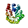
- Structure visualization
| Structure viewer | Molecule: MolmilJmol/JSmol |
|---|
- Downloads & links
-Download
| PDBx/mmCIF format | 1dl3.cif.gz | 87.8 KB | Display | PDBx/mmCIF format |
|---|---|---|---|---|
| PDB format | pdb1dl3.ent.gz | 67.5 KB | Display | PDB format |
| PDBx/mmJSON format | 1dl3.json.gz | Tree view | PDBx/mmJSON format | |
| Others |  Other downloads Other downloads |
-Validation report
| Arichive directory | https://data.pdbj.org/pub/pdb/validation_reports/dl/1dl3ftp://data.pdbj.org/pub/pdb/validation_reports/dl/1dl3 | HTTPS FTP |
|---|
-Related structure data
| Related structure data | |
|---|---|
| Similar structure data |
-Links
 PDBj
PDBj
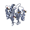
- Assembly
| Deposited unit | 
| ||||||||
|---|---|---|---|---|---|---|---|---|---|
| 1 | 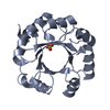
| ||||||||
| 2 | 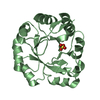
| ||||||||
| Unit cell |
|
-Components
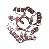
| #1: Protein | Mass: 22973.348 Da / Num. of mol.: 2 / Mutation: A25Y,F55E,I101W Source method: isolated from a genetically manipulated source Source: (gene. exp.) Thermotoga maritima (bacteria) / Plasmid: PET11C-TTRPF / Production host: Keywords: ENGINEERED MUTATIONReferences: UniProt: Q56320, phosphoribosylanthranilate isomerase #2: Chemical |   Mass: 96.063 Da / Num. of mol.: 2 / Source method: obtained synthetically / Formula: SO4 Mass: 96.063 Da / Num. of mol.: 2 / Source method: obtained synthetically / Formula: SO4#3: Water | ChemComp-HOH / |  Mass: 18.015 Da / Num. of mol.: 32 / Source method: isolated from a natural source / Formula: H2O Mass: 18.015 Da / Num. of mol.: 32 / Source method: isolated from a natural source / Formula: H2O |
|---|
-Experimental details
-Experiment
| Experiment | Method: X-RAY DIFFRACTION / Number of used crystals: 1 |
|---|
- Sample preparation
| Crystal | Density Matthews: 2.16 Å3/Da / Density % sol: 43.07 % | ||||||||||||||||||||||||||||||||||||||||
|---|---|---|---|---|---|---|---|---|---|---|---|---|---|---|---|---|---|---|---|---|---|---|---|---|---|---|---|---|---|---|---|---|---|---|---|---|---|---|---|---|---|
| Crystal grow | Method: vapor diffusion, hanging drop / pH: 7.5 Details: 1 MM EDTA 0.5 MM DTT 0.1 M HEPES/NAOH 16.7 MG/ML PROTEIN 0.75 M POTASSIUM DIHYDROGEN PHOSPHATE 0.75 M SODIUM DIHYDROGEN PHOSPHATE, pH 7.5, VAPOR DIFFUSION, HANGING DROP | ||||||||||||||||||||||||||||||||||||||||
| Crystal grow | *PLUS Method: vapor diffusion | ||||||||||||||||||||||||||||||||||||||||
| Components of the solutions | *PLUS
|
-Data collection
| Diffraction | Mean temperature: 298 K |
|---|---|
| Diffraction source | Source: ROTATING ANODE / Type: ENRAF-NONIUS FR571 / Wavelength: 1.54 |
| Detector | Type: MARRESEARCH / Detector: IMAGE PLATE / Date: May 18, 1998 |
| Radiation | Protocol: SINGLE WAVELENGTH / Monochromatic (M) / Laue (L): M / Scattering type: x-ray |
| Radiation wavelength | Wavelength: 1.54 Å / Relative weight: 1 |
| Reflection | Resolution: 2.7→20 Å / Num. all: 51809 / Num. obs: 11375 / % possible obs: 99.2 % / Observed criterion σ(F): 0 / Observed criterion σ(I): 0 / Redundancy: 4 % / Biso Wilson estimate: 31 Å2 / Rmerge(I) obs: 0.076 / Net I/σ(I): 8.2 |
| Reflection | *PLUS Num. measured all: 51809 |
| Reflection shell | *PLUS Highest resolution: 2.7 Å / Lowest resolution: 2.8 Å / % possible obs: 98.7 % / Rmerge(I) obs: 0.317 |
- Processing
| Software |
| |||||||||||||||||||||||||
|---|---|---|---|---|---|---|---|---|---|---|---|---|---|---|---|---|---|---|---|---|---|---|---|---|---|---|
| Refinement | Resolution: 2.7→20 Å / σ(F): 0 / σ(I): 0 / Stereochemistry target values: ENGH & HUBER
| |||||||||||||||||||||||||
| Refinement step | Cycle: LAST / Resolution: 2.7→20 Å
| |||||||||||||||||||||||||
| Software | *PLUS Name: X-PLOR / Version: 3.1 / Classification: refinement | |||||||||||||||||||||||||
| Refinement | *PLUS Highest resolution: 2.7 Å / Lowest resolution: 20 Å / σ(F): 0 / Rfactor obs: 0.175 | |||||||||||||||||||||||||
| Solvent computation | *PLUS | |||||||||||||||||||||||||
| Displacement parameters | *PLUS | |||||||||||||||||||||||||
| Refine LS restraints | *PLUS
|
