 Movie
Movie Controller


+ Open data

- Basic information
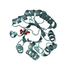
| Entry | Database: PDB / ID: 5bvl | ||||||
|---|---|---|---|---|---|---|---|
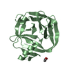
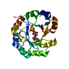
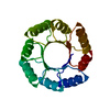
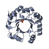
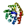
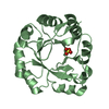
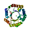
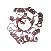
| Title | Crystal structure of a de novo designed TIM-barrel | ||||||
 Components Components | designed TIM barrel sTIM11 | ||||||
 Keywords Keywords | DE NOVO PROTEIN / TIM-barrel / computational design / idealized scaffold | ||||||
| Biological species | synthetic construct (others) | ||||||
| Method |  X-RAY DIFFRACTION / SYNCHROTRON / MOLECULAR REPLACEMENT / molecular replacement / Resolution: 1.992 Å X-RAY DIFFRACTION / SYNCHROTRON / MOLECULAR REPLACEMENT / molecular replacement / Resolution: 1.992 Å | ||||||
 Authors Authors | Feldmeier, K. / Hocker, B. | ||||||
 Citation Citation | Journal: Nat.Chem.Biol. / Year: 2016 Title: De novo design of a four-fold symmetric TIM-barrel protein with atomic-level accuracy. Authors: Huang, P.S. / Feldmeier, K. / Parmeggiani, F. / Fernandez Velasco, D.A. / Hocker, B. / Baker, D. | ||||||
| History |
|
- Structure visualization
| Structure viewer | Molecule:  MolmilJmol/JSmol MolmilJmol/JSmol |
|---|
- Downloads & links
-Download
| PDBx/mmCIF format | 5bvl.cif.gz | 78.6 KB | Display | PDBx/mmCIF format |
|---|---|---|---|---|
| PDB format | pdb5bvl.ent.gz | 59.6 KB | Display | PDB format |
| PDBx/mmJSON format | 5bvl.json.gz | Tree view | PDBx/mmJSON format | |
| Others |  Other downloads Other downloads |
-Validation report
| Arichive directory | https://data.pdbj.org/pub/pdb/validation_reports/bv/5bvlftp://data.pdbj.org/pub/pdb/validation_reports/bv/5bvl | HTTPS FTP |
|---|
-Related structure data
| Similar structure data |
|---|

-Links
 PDBj
PDBj
- Assembly
| Deposited unit | 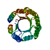
| ||||||||
|---|---|---|---|---|---|---|---|---|---|
| 1 |
| ||||||||
| Unit cell |
|
-Components
| #1: Protein | Mass: 22896.449 Da / Num. of mol.: 1 Source method: isolated from a genetically manipulated source Source: (gene. exp.) synthetic construct (others) / Production host:  |
|---|---|
| #2: Water | ChemComp-HOH /  Mass: 18.015 Da / Num. of mol.: 20 / Source method: isolated from a natural source / Formula: H2O Mass: 18.015 Da / Num. of mol.: 20 / Source method: isolated from a natural source / Formula: H2O |
-Experimental details
-Experiment
| Experiment | Method: X-RAY DIFFRACTION / Number of used crystals: 1 |
|---|
- Sample preparation
| Crystal | Density Matthews: 1.8 Å3/Da / Density % sol: 31.7 % / Description: Needle |
|---|---|
| Crystal grow | Temperature: 293 K / Method: vapor diffusion / pH: 5.5 Details: 25% PEG 3350, 0.2M magnesium chloride hexahydrate, 0.1M bis-tris |
-Data collection
| Diffraction | Mean temperature: 100 K |
|---|---|
| Diffraction source | Source: SYNCHROTRON / Site: SLS  / Beamline: X10SA / Wavelength: 1 Å / Beamline: X10SA / Wavelength: 1 Å |
| Detector | Type: DECTRIS PILATUS 6M / Detector: PIXEL / Date: Nov 2, 2014 |
| Radiation | Monochromator: DOUBLE-CRYSTAL SI / Protocol: SINGLE WAVELENGTH / Monochromatic (M) / Laue (L): M / Scattering type: x-ray |
| Radiation wavelength | Wavelength: 1 Å / Relative weight: 1 |
| Reflection | Resolution: 1.992→46.79 Å / Num. obs: 11894 / % possible obs: 98.26 % / Redundancy: 5.86 % / Biso Wilson estimate: 41.84 Å2 / Rmerge(I) obs: 0.05804 / Net I/σ(I): 15.24 |
| Reflection shell | Resolution: 1.992→2.064 Å / Rmerge(I) obs: 0.6607 / Mean I/σ(I) obs: 1.79 / % possible all: 93.2 |
-Phasing
| Phasing | Method: molecular replacement |
|---|
- Processing
| Software |
| |||||||||||||||||||||||||||||||||||
|---|---|---|---|---|---|---|---|---|---|---|---|---|---|---|---|---|---|---|---|---|---|---|---|---|---|---|---|---|---|---|---|---|---|---|---|---|
| Refinement | Method to determine structure: MOLECULAR REPLACEMENT Starting model: Rosetta backbone model Resolution: 1.992→46.79 Å / SU ML: 0.19 / Cross valid method: FREE R-VALUE / σ(F): 1.37 / Phase error: 27.25 / Stereochemistry target values: ML
| |||||||||||||||||||||||||||||||||||
| Solvent computation | Shrinkage radii: 0.9 Å / VDW probe radii: 1.11 Å / Solvent model: FLAT BULK SOLVENT MODEL | |||||||||||||||||||||||||||||||||||
| Displacement parameters | Biso mean: 61.9 Å2 | |||||||||||||||||||||||||||||||||||
| Refinement step | Cycle: LAST / Resolution: 1.992→46.79 Å
| |||||||||||||||||||||||||||||||||||
| Refine LS restraints |
| |||||||||||||||||||||||||||||||||||
| LS refinement shell |
|
