 Movie
Movie Controller

[English] 日本語
 Yorodumi
- PDB-1nsj: CRYSTAL STRUCTURE OF PHOSPHORIBOSYL ANTHRANILATE ISOMERASE FROM T... -
+ Open data

- Basic information
| Entry | Database: PDB / ID: 1nsj | ||||||
|---|---|---|---|---|---|---|---|
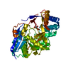
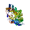
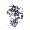
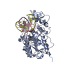
| Title | CRYSTAL STRUCTURE OF PHOSPHORIBOSYL ANTHRANILATE ISOMERASE FROM THERMOTOGA MARITIMA | ||||||
 Components Components | PHOSPHORIBOSYL ANTHRANILATE ISOMERASE | ||||||
 Keywords Keywords | ISOMERASE / PHOSPHORIBOSYL ANTHRANILATE ISOMERASE / THERMOSTABILITY | ||||||
| Function / homology |  Function and homology information Function and homology informationphosphoribosylanthranilate isomerase / phosphoribosylanthranilate isomerase activity / L-tryptophan biosynthetic process Similarity search - Function | ||||||
| Biological species |   Thermotoga maritima (bacteria) Thermotoga maritima (bacteria) | ||||||
| Method |  X-RAY DIFFRACTION / MIR / Resolution: 2 Å X-RAY DIFFRACTION / MIR / Resolution: 2 Å | ||||||
 Authors Authors | Hennig, M. / Jansonius, J.N.J. | ||||||
 Citation Citation | Journal: Biochemistry / Year: 1997 Title: Crystal structure at 2.0 A resolution of phosphoribosyl anthranilate isomerase from the hyperthermophile Thermotoga maritima: possible determinants of protein stability. Authors: Hennig, M. / Sterner, R. / Kirschner, K. / Jansonius, J.N. #1: Journal: Protein Sci. / Year: 1996Title: Phosphoribosyl Anthranilate Isomerase from Thermotoga Maritima is an Extremely Stable and Active Homodimer Authors: Sterner, R. / Kleemann, G.R. / Szadkowski, H. / Lustig, A. / Hennig, M. / Kirschner, K. | ||||||
| History |
|
- Structure visualization
| Structure viewer | Molecule: MolmilJmol/JSmol |
|---|
- Downloads & links
-Download
| PDBx/mmCIF format | 1nsj.cif.gz | 54.8 KB | Display | PDBx/mmCIF format |
|---|---|---|---|---|
| PDB format | pdb1nsj.ent.gz | 40 KB | Display | PDB format |
| PDBx/mmJSON format | 1nsj.json.gz | Tree view | PDBx/mmJSON format | |
| Others |  Other downloads Other downloads |
-Validation report
| Arichive directory | https://data.pdbj.org/pub/pdb/validation_reports/ns/1nsjftp://data.pdbj.org/pub/pdb/validation_reports/ns/1nsj | HTTPS FTP |
|---|
-Related structure data
| Similar structure data |
|---|


-Links
 PDBj
PDBj
- Assembly
| Deposited unit | 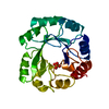
| ||||||||
|---|---|---|---|---|---|---|---|---|---|
| 1 | 
| ||||||||
| Unit cell |
|
-Components
| #1: Protein | Mass: 23070.549 Da / Num. of mol.: 1 Source method: isolated from a genetically manipulated source Source: (gene. exp.) Thermotoga maritima (bacteria) / Gene: TRPF / Plasmid: PQE-60 / Gene (production host): TRPF / Production host: References: UniProt: Q56320, phosphoribosylanthranilate isomerase |
|---|---|
| #2: Chemical | ChemComp-PO4 /   Mass: 94.971 Da / Num. of mol.: 1 / Source method: obtained synthetically / Formula: PO4 Mass: 94.971 Da / Num. of mol.: 1 / Source method: obtained synthetically / Formula: PO4 |
| #3: Water | ChemComp-HOH /  Mass: 18.015 Da / Num. of mol.: 106 / Source method: isolated from a natural source / Formula: H2O Mass: 18.015 Da / Num. of mol.: 106 / Source method: isolated from a natural source / Formula: H2O |
-Experimental details
-Experiment
| Experiment | Method: X-RAY DIFFRACTION / Number of used crystals: 3 |
|---|
- Sample preparation
| Crystal | Density Matthews: 2.71 Å3/Da / Density % sol: 45 % / Description: HEAVY ATOM DERIVATIVE PHASING | ||||||||||||||||||||||||||||||||||||||||||||||||
|---|---|---|---|---|---|---|---|---|---|---|---|---|---|---|---|---|---|---|---|---|---|---|---|---|---|---|---|---|---|---|---|---|---|---|---|---|---|---|---|---|---|---|---|---|---|---|---|---|---|
| Crystal grow | pH: 7.5 Details: PROTEIN 19.4 MG/ML IN 0.05 M POTASSIUM PHOSPHATE PH 7.5, 1MM EDTA, 0.4 MM DTT MIXED WITH RESERVOIR 1:1 CONTAINING 1.5-2.1 M AMMONIUM SULFATE, 10 MM COCL2, 0.1 M MES PH 6.5, PH range: 6.5-7.5 | ||||||||||||||||||||||||||||||||||||||||||||||||
| Crystal grow | *PLUS Temperature: 4 ℃ / Method: vapor diffusion, hanging dropDetails: drop solution was mixed with an equal volume of reservoir solution | ||||||||||||||||||||||||||||||||||||||||||||||||
| Components of the solutions | *PLUS
|
-Data collection
| Diffraction | Mean temperature: 277 K |
|---|---|
| Diffraction source | Source: ROTATING ANODE / Type: ELLIOTT GX-18 / Wavelength: 1.5418 |
| Detector | Type: MARRESEARCH / Detector: IMAGE PLATE / Date: Jan 9, 1995 |
| Radiation | Monochromator: GRAPHITE(002) / Monochromatic (M) / Laue (L): M / Scattering type: x-ray |
| Radiation wavelength | Wavelength: 1.5418 Å / Relative weight: 1 |
| Reflection | Resolution: 2→20 Å / Num. obs: 17903 / % possible obs: 98.8 % / Observed criterion σ(I): 0 / Redundancy: 6.6 % / Rmerge(I) obs: 0.064 / Net I/σ(I): 8.6 |
| Reflection shell | Resolution: 2→2.11 Å / Redundancy: 4.5 % / Rmerge(I) obs: 0.311 / Mean I/σ(I) obs: 2.4 / % possible all: 96.2 |
| Reflection | *PLUS Num. measured all: 118018 |
| Reflection shell | *PLUS % possible obs: 96.2 % |
- Processing
| Software |
| ||||||||||||||||||||||||||||||||||||||||||||||||||||||||||||
|---|---|---|---|---|---|---|---|---|---|---|---|---|---|---|---|---|---|---|---|---|---|---|---|---|---|---|---|---|---|---|---|---|---|---|---|---|---|---|---|---|---|---|---|---|---|---|---|---|---|---|---|---|---|---|---|---|---|---|---|---|---|
| Refinement | Method to determine structure: MIR / Resolution: 2→15 Å / σ(F): 0 Details: RESIDUES 129 - 136 ARE IN THE MODEL WITH OCCUPANCIES = 0, BECAUSE THERE IS NO ELECTRON DENSITY FOR THESE RESIDUES; THEY WERE BUILT STEREOCHEMICALLY ONLY.
| ||||||||||||||||||||||||||||||||||||||||||||||||||||||||||||
| Displacement parameters | Biso mean: 32.1 Å2 | ||||||||||||||||||||||||||||||||||||||||||||||||||||||||||||
| Refinement step | Cycle: LAST / Resolution: 2→15 Å
| ||||||||||||||||||||||||||||||||||||||||||||||||||||||||||||
| Refine LS restraints |
| ||||||||||||||||||||||||||||||||||||||||||||||||||||||||||||
| LS refinement shell | Resolution: 2→2.03 Å / Total num. of bins used: 20
| ||||||||||||||||||||||||||||||||||||||||||||||||||||||||||||
| Software | *PLUS Name: X-PLOR / Classification: refinement | ||||||||||||||||||||||||||||||||||||||||||||||||||||||||||||
| Refinement | *PLUS Lowest resolution: 10 Å / % reflection Rfree: 5 % | ||||||||||||||||||||||||||||||||||||||||||||||||||||||||||||
| Solvent computation | *PLUS | ||||||||||||||||||||||||||||||||||||||||||||||||||||||||||||
| Displacement parameters | *PLUS |
