 Movie
Movie Controller

[English] 日本語
 Yorodumi
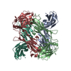
- PDB-3djd: Crystal structure of the deglycating enzyme fructosamine oxidase ... -
+ Open data

- Basic information
| Entry | Database: PDB / ID: 3djd | ||||||
|---|---|---|---|---|---|---|---|
| Title | Crystal structure of the deglycating enzyme fructosamine oxidase from Aspergillus fumigatus (Amadoriase II) | ||||||
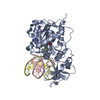
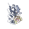
 Components Components | Fructosyl amine: oxygen oxidoreductase | ||||||
 Keywords Keywords | OXIDOREDUCTASE / fructosyl-amino acid / amadoriase / deglycation / fructosamine oxidase | ||||||
| Function / homology |  Function and homology information Function and homology informationsaccharopine oxidase activity / sarcosine oxidase activity / flavin adenine dinucleotide binding Similarity search - Function | ||||||
| Biological species |  | ||||||
| Method |  X-RAY DIFFRACTION / SYNCHROTRON / SAD / Resolution: 1.75 Å X-RAY DIFFRACTION / SYNCHROTRON / SAD / Resolution: 1.75 Å | ||||||
 Authors Authors | Collard, F. / Zhang, J. / Nemet, I. / Qanungo, K.R. / Monnier, V.M. / Yee, V.C. | ||||||
 Citation Citation | Journal: To be Published Title: Crystal structure of the deglycating enzyme fructosamine oxidase (FAOX-II) Authors: Collard, F. / Zhang, J. / Nemet, I. / Qanungo, K.R. / Monnier, V.M. / Yee, V.C. | ||||||
| History |
|
- Structure visualization
| Structure viewer | Molecule: MolmilJmol/JSmol |
|---|
- Downloads & links
-Download
| PDBx/mmCIF format | 3djd.cif.gz | 201.8 KB | Display | PDBx/mmCIF format |
|---|---|---|---|---|
| PDB format | pdb3djd.ent.gz | 158.9 KB | Display | PDB format |
| PDBx/mmJSON format | 3djd.json.gz | Tree view | PDBx/mmJSON format | |
| Others |  Other downloads Other downloads |
-Validation report
| Arichive directory | https://data.pdbj.org/pub/pdb/validation_reports/dj/3djdftp://data.pdbj.org/pub/pdb/validation_reports/dj/3djd | HTTPS FTP |
|---|
-Related structure data


-Links
 PDBj
PDBj- Assembly
| Deposited unit | 
| ||||||||
|---|---|---|---|---|---|---|---|---|---|
| 1 | 
| ||||||||
| 2 | 
| ||||||||
| Unit cell |
|
-Components
| #1: Protein | Mass: 49429.609 Da / Num. of mol.: 2 Source method: isolated from a genetically manipulated source Source: (gene. exp.)  #2: Chemical |   Mass: 785.550 Da / Num. of mol.: 2 / Source method: obtained synthetically / Formula: C27H33N9O15P2 / Comment: FAD*YM Mass: 785.550 Da / Num. of mol.: 2 / Source method: obtained synthetically / Formula: C27H33N9O15P2 / Comment: FAD*YM#3: Water | ChemComp-HOH / |  Mass: 18.015 Da / Num. of mol.: 842 / Source method: isolated from a natural source / Formula: H2O Mass: 18.015 Da / Num. of mol.: 842 / Source method: isolated from a natural source / Formula: H2OHas protein modification | Y | Sequence details | ACCORDING TO THE AUTHORS THE SEQUENCE WAS VERIFIED BY SEQUENCING AND DIFFERS FROM THE UNP SEQUENCE ...ACCORDING TO THE AUTHORS THE SEQUENCE WAS VERIFIED BY SEQUENCING | |
|---|
-Experimental details
-Experiment
| Experiment | Method: X-RAY DIFFRACTION / Number of used crystals: 1 |
|---|
- Sample preparation
| Crystal | Density Matthews: 2.16 Å3/Da / Density % sol: 42.94 % |
|---|---|
| Crystal grow | Temperature: 293 K / Method: vapor diffusion, sitting drop / pH: 7.4 Details: 0.1 M Hepes pH 7.4, 10 % iso-propanol, 18 % PEG 4000, protein solution 15 mg/ml, VAPOR DIFFUSION, SITTING DROP, temperature 293K |
-Data collection
| Diffraction | Mean temperature: 100 K |
|---|---|
| Diffraction source | Source: SYNCHROTRON / Site: APS  / Beamline: 19-ID / Wavelength: 0.97929 Å / Beamline: 19-ID / Wavelength: 0.97929 Å |
| Detector | Type: ADSC QUANTUM 315 / Detector: CCD / Date: Aug 7, 2006 |
| Radiation | Protocol: SINGLE WAVELENGTH / Monochromatic (M) / Laue (L): M / Scattering type: x-ray |
| Radiation wavelength | Wavelength: 0.97929 Å / Relative weight: 1 |
| Reflection | Resolution: 1.75→36.96 Å / Num. obs: 85107 / % possible obs: 99.9 % / Observed criterion σ(F): 0 / Observed criterion σ(I): 0 / Redundancy: 13.3 % / Rmerge(I) obs: 0.087 / Net I/σ(I): 37.4 |
| Reflection shell | Resolution: 1.75→1.81 Å / Redundancy: 12 % / Rmerge(I) obs: 0.623 / Mean I/σ(I) obs: 3.9 / % possible all: 99.6 |
- Processing
| Software |
| ||||||||||||||||||||||||||||||||||||||||||||||||||||||||||||||||||||||||||||||||||||||||||||||||||||||||||||||||||||||||||||||||||||||||||||||||||||||||||||||||||||||||||
|---|---|---|---|---|---|---|---|---|---|---|---|---|---|---|---|---|---|---|---|---|---|---|---|---|---|---|---|---|---|---|---|---|---|---|---|---|---|---|---|---|---|---|---|---|---|---|---|---|---|---|---|---|---|---|---|---|---|---|---|---|---|---|---|---|---|---|---|---|---|---|---|---|---|---|---|---|---|---|---|---|---|---|---|---|---|---|---|---|---|---|---|---|---|---|---|---|---|---|---|---|---|---|---|---|---|---|---|---|---|---|---|---|---|---|---|---|---|---|---|---|---|---|---|---|---|---|---|---|---|---|---|---|---|---|---|---|---|---|---|---|---|---|---|---|---|---|---|---|---|---|---|---|---|---|---|---|---|---|---|---|---|---|---|---|---|---|---|---|---|---|---|
| Refinement | Method to determine structure: SAD / Resolution: 1.75→36.96 Å / Cor.coef. Fo:Fc: 0.96 / Cor.coef. Fo:Fc free: 0.939 / SU B: 2.168 / SU ML: 0.071 / Cross valid method: THROUGHOUT / ESU R: 0.116 / ESU R Free: 0.112 / Stereochemistry target values: MAXIMUM LIKELIHOOD / Details: HYDROGENS HAVE BEEN ADDED IN THE RIDING POSITIONS
| ||||||||||||||||||||||||||||||||||||||||||||||||||||||||||||||||||||||||||||||||||||||||||||||||||||||||||||||||||||||||||||||||||||||||||||||||||||||||||||||||||||||||||
| Solvent computation | Ion probe radii: 0.8 Å / Shrinkage radii: 0.8 Å / VDW probe radii: 1.4 Å / Solvent model: MASK | ||||||||||||||||||||||||||||||||||||||||||||||||||||||||||||||||||||||||||||||||||||||||||||||||||||||||||||||||||||||||||||||||||||||||||||||||||||||||||||||||||||||||||
| Displacement parameters | Biso mean: 18.925 Å2
| ||||||||||||||||||||||||||||||||||||||||||||||||||||||||||||||||||||||||||||||||||||||||||||||||||||||||||||||||||||||||||||||||||||||||||||||||||||||||||||||||||||||||||
| Refine analyze |
| ||||||||||||||||||||||||||||||||||||||||||||||||||||||||||||||||||||||||||||||||||||||||||||||||||||||||||||||||||||||||||||||||||||||||||||||||||||||||||||||||||||||||||
| Refinement step | Cycle: LAST / Resolution: 1.75→36.96 Å
| ||||||||||||||||||||||||||||||||||||||||||||||||||||||||||||||||||||||||||||||||||||||||||||||||||||||||||||||||||||||||||||||||||||||||||||||||||||||||||||||||||||||||||
| Refine LS restraints |
| ||||||||||||||||||||||||||||||||||||||||||||||||||||||||||||||||||||||||||||||||||||||||||||||||||||||||||||||||||||||||||||||||||||||||||||||||||||||||||||||||||||||||||
| LS refinement shell | Resolution: 1.75→1.796 Å / Total num. of bins used: 20
|
