- PDB-1d9g: BOVINE INTERFERON-GAMMA AT 2.9 ANGSTROMS -
+
Open data
ID or keywords:
Loading...
-
Basic information
Entry
Database: PDB / ID: 1d9g
Title
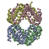
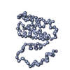
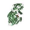
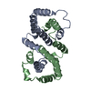
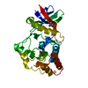
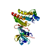
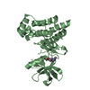
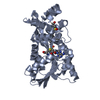
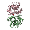
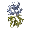
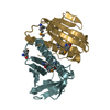
BOVINE INTERFERON-GAMMA AT 2.9 ANGSTROMS
Components
INTERFERON-GAMMA
Keywords
IMMUNE SYSTEM / helical homodimer
Function / homology
Function and homology information
Interferon gamma signaling / Regulation of IFNG signaling / IFNG signaling activates MAPKs / positive regulation of fructose 1,6-bisphosphate metabolic process / positive regulation of tumor necrosis factor (ligand) superfamily member 11 production / type II interferon receptor binding / : / positive regulation of vitamin D biosynthetic process / positive regulation of interleukin-23 production / type III interferon-mediated signaling pathway ...Interferon gamma signaling / Regulation of IFNG signaling / IFNG signaling activates MAPKs / positive regulation of fructose 1,6-bisphosphate metabolic process / positive regulation of tumor necrosis factor (ligand) superfamily member 11 production / type II interferon receptor binding / : / positive regulation of vitamin D biosynthetic process / positive regulation of interleukin-23 production / type III interferon-mediated signaling pathway / positive regulation of smooth muscle cell apoptotic process / negative regulation of interleukin-17 production / positive regulation of osteoclast differentiation / macrophage activation involved in immune response / positive regulation of membrane protein ectodomain proteolysis / positive regulation of neurogenesis / positive regulation of amyloid-beta formation / interleukin-2-mediated signaling pathway / Fc-gamma receptor signaling pathway involved in phagocytosis / regulation of insulin secretion / humoral immune response / macrophage differentiation / positive regulation of epithelial cell migration / type II interferon-mediated signaling pathway / cell surface receptor signaling pathway via JAK-STAT / extrinsic apoptotic signaling pathway / positive regulation of interleukin-12 production / positive regulation of B cell proliferation / positive regulation of chemokine production / positive regulation of autophagy / astrocyte activation / cytokine activity / negative regulation of smooth muscle cell proliferation / microglial cell activation / positive regulation of protein-containing complex assembly / positive regulation of T cell cytokine production / positive regulation of protein import into nucleus / positive regulation of interleukin-6 production / positive regulation of nitric oxide biosynthetic process / positive regulation of inflammatory response / defense response to virus / adaptive immune response / negative regulation of DNA-templated transcription / positive regulation of transcription by RNA polymerase II / : Similarity search - Function
In the structure databanks used in Yorodumi, some data are registered as the other names, "COVID-19 virus" and "2019-nCoV". Here are the details of the virus and the list of structure data.
Jan 31, 2019. EMDB accession codes are about to change! (news from PDBe EMDB page)
EMDB accession codes are about to change! (news from PDBe EMDB page)
The allocation of 4 digits for EMDB accession codes will soon come to an end. Whilst these codes will remain in use, new EMDB accession codes will include an additional digit and will expand incrementally as the available range of codes is exhausted. The current 4-digit format prefixed with “EMD-” (i.e. EMD-XXXX) will advance to a 5-digit format (i.e. EMD-XXXXX), and so on. It is currently estimated that the 4-digit codes will be depleted around Spring 2019, at which point the 5-digit format will come into force.
The EM Navigator/Yorodumi systems omit the EMD- prefix.
Related info.:Q: What is EMD? / ID/Accession-code notation in Yorodumi/EM Navigator
Yorodumi is a browser for structure data from EMDB, PDB, SASBDB, etc.
This page is also the successor to EM Navigator detail page, and also detail information page/front-end page for Omokage search.
The word "yorodu" (or yorozu) is an old Japanese word meaning "ten thousand". "mi" (miru) is to see.
Related info.:EMDB / PDB / SASBDB / Comparison of 3 databanks / Yorodumi Search / Aug 31, 2016. New EM Navigator & Yorodumi / Yorodumi Papers / Jmol/JSmol / Function and homology information / Changes in new EM Navigator and Yorodumi
 Movie
Movie Controller


 Open data
 Basic information
 Components
 Keywords
 Function and homology information
 X-RAY DIFFRACTION / Resolution: 2.9 Å
  Authors
 Citation
 Structure visualization
 Downloads & links
 Other downloads

 PDBj
PDBj


 Assembly
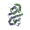

 Sample preparation
 Processing