 Movie
Movie Controller

[English] 日本語
 Yorodumi
- PDB-1d4f: CRYSTAL STRUCTURE OF RECOMBINANT RAT-LIVER D244E MUTANT S-ADENOSY... -
+ Open data

- Basic information
| Entry | Database: PDB / ID: 1d4f | ||||||
|---|---|---|---|---|---|---|---|
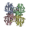
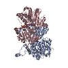
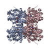
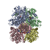
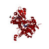
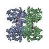
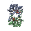
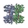
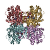
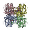
| Title | CRYSTAL STRUCTURE OF RECOMBINANT RAT-LIVER D244E MUTANT S-ADENOSYLHOMOCYSTEINE HYDROLASE | ||||||
 Components Components | S-ADENOSYLHOMOCYSTEINE HYDROLASE | ||||||
 Keywords Keywords | HYDROLASE / S-adenosylhomocysteine hydrolase / AdoHcyase / AdoHcy / Mutagenesis / X-ray crystal structure / Enzyme structure | ||||||
| Function / homology |  Function and homology information Function and homology informationadenosylselenohomocysteinase activity / : / Sulfur amino acid metabolism / S-methylmethionine cycle / circadian sleep/wake cycle / adenyl nucleotide binding / chronic inflammatory response to antigenic stimulus / Methylation / adenosylhomocysteinase / adenosylhomocysteinase activity ...adenosylselenohomocysteinase activity / : / Sulfur amino acid metabolism / S-methylmethionine cycle / circadian sleep/wake cycle / adenyl nucleotide binding / chronic inflammatory response to antigenic stimulus / Methylation / adenosylhomocysteinase / adenosylhomocysteinase activity / L-methionine cycle / one-carbon metabolic process / response to nutrient / NAD binding / melanosome / response to hypoxia / copper ion binding / endoplasmic reticulum / identical protein binding / nucleus / cytosol Similarity search - Function | ||||||
| Biological species |  | ||||||
| Method |  X-RAY DIFFRACTION / Resolution: 2.8 Å X-RAY DIFFRACTION / Resolution: 2.8 Å | ||||||
 Authors Authors | Komoto, J. / Huang, Y. / Takusagawa, F. / Gomi, T. / Ogawa, H. / Takata, Y. / Fujioka, M. | ||||||
 Citation Citation | Journal: J.Biol.Chem. / Year: 2000 Title: Effects of site-directed mutagenesis on structure and function of recombinant rat liver S-adenosylhomocysteine hydrolase. Crystal structure of D244E mutant enzyme. Authors: Komoto, J. / Huang, Y. / Gomi, T. / Ogawa, H. / Takata, Y. / Fujioka, M. / Takusagawa, F. | ||||||
| History |
|
- Structure visualization
| Structure viewer | Molecule: MolmilJmol/JSmol |
|---|
- Downloads & links
-Download
| PDBx/mmCIF format | 1d4f.cif.gz | 342.4 KB | Display | PDBx/mmCIF format |
|---|---|---|---|---|
| PDB format | pdb1d4f.ent.gz | 278.9 KB | Display | PDB format |
| PDBx/mmJSON format | 1d4f.json.gz | Tree view | PDBx/mmJSON format | |
| Others |  Other downloads Other downloads |
-Validation report
| Arichive directory | https://data.pdbj.org/pub/pdb/validation_reports/d4/1d4fftp://data.pdbj.org/pub/pdb/validation_reports/d4/1d4f | HTTPS FTP |
|---|
-Related structure data
| Related structure data | |
|---|---|
| Similar structure data |
-Links
 PDBj
PDBj
- Assembly
| Deposited unit | 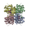
| ||||||||||||
|---|---|---|---|---|---|---|---|---|---|---|---|---|---|
| 1 |
| ||||||||||||
| Unit cell |
| ||||||||||||
| Components on special symmetry positions |
|
-Components
| #1: Protein | Mass: 47479.738 Da / Num. of mol.: 4 / Mutation: D244E Source method: isolated from a genetically manipulated source Source: (gene. exp.)  #2: Chemical | ChemComp-NAD /   Mass: 663.425 Da / Num. of mol.: 4 / Source method: obtained synthetically / Formula: C21H27N7O14P2 / Comment: NAD*YM Mass: 663.425 Da / Num. of mol.: 4 / Source method: obtained synthetically / Formula: C21H27N7O14P2 / Comment: NAD*YM#3: Chemical | ChemComp-ADN /   Mass: 267.241 Da / Num. of mol.: 4 / Source method: obtained synthetically / Formula: C10H13N5O4 Mass: 267.241 Da / Num. of mol.: 4 / Source method: obtained synthetically / Formula: C10H13N5O4#4: Water | ChemComp-HOH / |  Mass: 18.015 Da / Num. of mol.: 473 / Source method: isolated from a natural source / Formula: H2O Mass: 18.015 Da / Num. of mol.: 473 / Source method: isolated from a natural source / Formula: H2O |
|---|
-Experimental details
-Experiment
| Experiment | Method: X-RAY DIFFRACTION / Number of used crystals: 1 |
|---|
- Sample preparation
| Crystal | Density Matthews: 2.44 Å3/Da / Density % sol: 49.51 % | |||||||||||||||||||||||||||||||||||
|---|---|---|---|---|---|---|---|---|---|---|---|---|---|---|---|---|---|---|---|---|---|---|---|---|---|---|---|---|---|---|---|---|---|---|---|---|
| Crystal grow | Temperature: 295 K / Method: vapor diffusion, hanging drop / pH: 7.2 Details: 22% PEG 4000, 50 mM Tris/HCl, 2% glycerol, 10% isopropanol, and 1 mM DTT. Protein concentration is 10 mg/mL., pH 7.2, VAPOR DIFFUSION, HANGING DROP, temperature 295K | |||||||||||||||||||||||||||||||||||
| Crystal grow | *PLUS Temperature: 22 ℃ / Details: used seeding | |||||||||||||||||||||||||||||||||||
| Components of the solutions | *PLUS
|
-Data collection
| Diffraction | Mean temperature: 93 K |
|---|---|
| Diffraction source | Source: ROTATING ANODE / Type: RIGAKU RU200 / Wavelength: 1.5418 |
| Detector | Type: RIGAKU RAXIS IIC / Detector: IMAGE PLATE / Date: Nov 27, 1996 |
| Radiation | Protocol: SINGLE WAVELENGTH / Monochromatic (M) / Laue (L): M / Scattering type: x-ray |
| Radiation wavelength | Wavelength: 1.5418 Å / Relative weight: 1 |
| Reflection | Resolution: 2.8→8 Å / Num. all: 241990 / Num. obs: 241990 / % possible obs: 91.8 % / Observed criterion σ(F): 0 / Observed criterion σ(I): 0 / Redundancy: 4.7 % / Biso Wilson estimate: 15 Å2 / Rmerge(I) obs: 0.105 / Net I/σ(I): 24.4 |
| Reflection shell | Resolution: 2.8→2.9 Å / Redundancy: 2.7 % / Rmerge(I) obs: 0.265 / Num. unique all: 3808 / % possible all: 83.7 |
| Reflection | *PLUS Lowest resolution: 20 Å / Num. obs: 43000 / % possible obs: 85.6 % / Num. measured all: 241990 / Rmerge(I) obs: 0.093 |
- Processing
| Software |
| |||||||||||||||||||||||||
|---|---|---|---|---|---|---|---|---|---|---|---|---|---|---|---|---|---|---|---|---|---|---|---|---|---|---|
| Refinement | Resolution: 2.8→8 Å / Cross valid method: THROUGHOUT / σ(F): 0 / σ(I): 0 / Stereochemistry target values: Engh & Huber
| |||||||||||||||||||||||||
| Refinement step | Cycle: LAST / Resolution: 2.8→8 Å
| |||||||||||||||||||||||||
| Refine LS restraints |
| |||||||||||||||||||||||||
| Software | *PLUS Name: X-PLOR / Version: 3.843 / Classification: refinement | |||||||||||||||||||||||||
| Refinement | *PLUS Highest resolution: 2.8 Å / Lowest resolution: 8 Å / σ(F): 0 / % reflection Rfree: 10 % | |||||||||||||||||||||||||
| Solvent computation | *PLUS | |||||||||||||||||||||||||
| Displacement parameters | *PLUS | |||||||||||||||||||||||||
| Refine LS restraints | *PLUS
|
