 Movie
Movie Controller


+ Open data

- Basic information
| Entry | Database: PDB / ID: 1b3r | ||||||
|---|---|---|---|---|---|---|---|
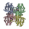
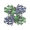
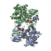
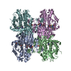
| Title | RAT LIVER S-ADENOSYLHOMOCYSTEIN HYDROLASE | ||||||
 Components Components | PROTEIN (S-ADENOSYLHOMOCYSTEINE HYDROLASE) | ||||||
 Keywords Keywords | HYDROLASE / ADEONSYLHOMOCYSTEINE / ADOHCY / ADOHCYASE | ||||||
| Function / homology |  Function and homology information Function and homology informationadenosylselenohomocysteinase activity / : / Sulfur amino acid metabolism / S-methylmethionine cycle / circadian sleep/wake cycle / adenyl nucleotide binding / chronic inflammatory response to antigenic stimulus / Methylation / adenosylhomocysteinase / adenosylhomocysteinase activity ...adenosylselenohomocysteinase activity / : / Sulfur amino acid metabolism / S-methylmethionine cycle / circadian sleep/wake cycle / adenyl nucleotide binding / chronic inflammatory response to antigenic stimulus / Methylation / adenosylhomocysteinase / adenosylhomocysteinase activity / L-methionine cycle / one-carbon metabolic process / response to nutrient / NAD binding / melanosome / response to hypoxia / copper ion binding / endoplasmic reticulum / identical protein binding / nucleus / cytosol Similarity search - Function | ||||||
| Biological species |  | ||||||
| Method |  X-RAY DIFFRACTION / MOLECULAR REPLACEMENT / Resolution: 2.8 Å X-RAY DIFFRACTION / MOLECULAR REPLACEMENT / Resolution: 2.8 Å | ||||||
 Authors Authors | Hu, Y. / Komoto, J. / Huang, Y. / Takusagawa, F. / Gomi, T. / Ogawa, H. / Takata, Y. / Fujioka, M. | ||||||
 Citation Citation | Journal: Biochemistry / Year: 1999 Title: Crystal structure of S-adenosylhomocysteine hydrolase from rat liver. Authors: Hu, Y. / Komoto, J. / Huang, Y. / Gomi, T. / Ogawa, H. / Takata, Y. / Fujioka, M. / Takusagawa, F. | ||||||
| History |
|
- Structure visualization
| Structure viewer | Molecule: MolmilJmol/JSmol |
|---|
- Downloads & links
-Download
| PDBx/mmCIF format | 1b3r.cif.gz | 409.3 KB | Display | PDBx/mmCIF format |
|---|---|---|---|---|
| PDB format | pdb1b3r.ent.gz | 338.8 KB | Display | PDB format |
| PDBx/mmJSON format | 1b3r.json.gz | Tree view | PDBx/mmJSON format | |
| Others |  Other downloads Other downloads |
-Validation report
| Arichive directory | https://data.pdbj.org/pub/pdb/validation_reports/b3/1b3rftp://data.pdbj.org/pub/pdb/validation_reports/b3/1b3r | HTTPS FTP |
|---|
-Related structure data
| Similar structure data |
|---|
-Links
 PDBj
PDBj- Assembly
| Deposited unit | 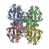
| ||||||||||||||||
|---|---|---|---|---|---|---|---|---|---|---|---|---|---|---|---|---|---|
| 1 |
| ||||||||||||||||
| Unit cell |
| ||||||||||||||||
| Noncrystallographic symmetry (NCS) | NCS oper:
|
-Components
| #1: Protein | Mass: 47465.711 Da / Num. of mol.: 4 / Fragment: CATALYTIC DOMAIN (1 - 181 & 352 - 402) Source method: isolated from a genetically manipulated source Details: EACH SUBUNIT CONTAINS ONE NAD+ MOLECULE. / Source: (gene. exp.)  #2: Chemical | ChemComp-NAD /   Mass: 663.425 Da / Num. of mol.: 4 / Source method: obtained synthetically / Formula: C21H27N7O14P2 / Comment: NAD*YM Mass: 663.425 Da / Num. of mol.: 4 / Source method: obtained synthetically / Formula: C21H27N7O14P2 / Comment: NAD*YM#3: Water | ChemComp-HOH / |  Mass: 18.015 Da / Num. of mol.: 413 / Source method: isolated from a natural source / Formula: H2O Mass: 18.015 Da / Num. of mol.: 413 / Source method: isolated from a natural source / Formula: H2OHas protein modification | N | |
|---|
-Experimental details
-Experiment
| Experiment | Method: X-RAY DIFFRACTION / Number of used crystals: 2 |
|---|
- Sample preparation
| Crystal | Density Matthews: 3.1 Å3/Da / Density % sol: 60 % | ||||||||||||||||||||||||||||||
|---|---|---|---|---|---|---|---|---|---|---|---|---|---|---|---|---|---|---|---|---|---|---|---|---|---|---|---|---|---|---|---|
| Crystal grow | pH: 6.8 Details: 50 MM TRIS/HCL (PH 6.8), 6 MM MGCL2, 5% MPD, 15% PEG-6K | ||||||||||||||||||||||||||||||
| Components of the solutions |
| ||||||||||||||||||||||||||||||
| Crystal | *PLUS | ||||||||||||||||||||||||||||||
| Crystal grow | *PLUS Temperature: 26 ℃ / Method: vapor diffusion, hanging drop | ||||||||||||||||||||||||||||||
| Components of the solutions | *PLUS
|
-Data collection
| Diffraction | Mean temperature: 93 K |
|---|---|
| Diffraction source | Source: ROTATING ANODE / Type: RIGAKU RU200 / Wavelength: 1.5418 |
| Detector | Type: RIGAKU / Detector: IMAGE PLATE / Date: Jan 1, 1998 |
| Radiation | Protocol: SINGLE WAVELENGTH / Monochromatic (M) / Laue (L): M / Scattering type: x-ray |
| Radiation wavelength | Wavelength: 1.5418 Å / Relative weight: 1 |
| Reflection | Resolution: 2.8→20 Å / Num. obs: 56314 / % possible obs: 97.5 % / Observed criterion σ(I): 0 / Redundancy: 5 % / Rmerge(I) obs: 0.07 / Rsym value: 0.07 / Net I/σ(I): 10.1 |
| Reflection shell | Resolution: 2.8→3 Å / Redundancy: 4.2 % / Rmerge(I) obs: 0.173 / Mean I/σ(I) obs: 3.4 / Rsym value: 0.173 / % possible all: 98 |
| Reflection | *PLUS Num. measured all: 280288 / Rmerge(I) obs: 0.07 |
- Processing
| Software |
| ||||||||||||||||||||||||||||||||||||||||||||||||||||||||||||
|---|---|---|---|---|---|---|---|---|---|---|---|---|---|---|---|---|---|---|---|---|---|---|---|---|---|---|---|---|---|---|---|---|---|---|---|---|---|---|---|---|---|---|---|---|---|---|---|---|---|---|---|---|---|---|---|---|---|---|---|---|---|
| Refinement | Method to determine structure: MOLECULAR REPLACEMENT Starting model: NAD-BINDING DOMAIN IN FORMATE DEHYDROGENASE Resolution: 2.8→8 Å / Data cutoff high absF: 0 / Data cutoff low absF: 0 / Cross valid method: APPLIED / σ(F): 0
| ||||||||||||||||||||||||||||||||||||||||||||||||||||||||||||
| Displacement parameters | Biso mean: 21.5 Å2 | ||||||||||||||||||||||||||||||||||||||||||||||||||||||||||||
| Refinement step | Cycle: LAST / Resolution: 2.8→8 Å
| ||||||||||||||||||||||||||||||||||||||||||||||||||||||||||||
| Refine LS restraints |
| ||||||||||||||||||||||||||||||||||||||||||||||||||||||||||||
| LS refinement shell | Resolution: 2.8→2.92 Å / Total num. of bins used: 8
| ||||||||||||||||||||||||||||||||||||||||||||||||||||||||||||
| Xplor file |
| ||||||||||||||||||||||||||||||||||||||||||||||||||||||||||||
| Software | *PLUS Name: X-PLOR / Version: 3.1 / Classification: refinement | ||||||||||||||||||||||||||||||||||||||||||||||||||||||||||||
| Refinement | *PLUS Highest resolution: 2.8 Å / Lowest resolution: 8 Å / σ(F): 0 / % reflection Rfree: 5 % / Rfactor obs: 0.197 | ||||||||||||||||||||||||||||||||||||||||||||||||||||||||||||
| Solvent computation | *PLUS | ||||||||||||||||||||||||||||||||||||||||||||||||||||||||||||
| Displacement parameters | *PLUS Biso mean: 21.5 Å2 | ||||||||||||||||||||||||||||||||||||||||||||||||||||||||||||
| Refine LS restraints | *PLUS
| ||||||||||||||||||||||||||||||||||||||||||||||||||||||||||||
| LS refinement shell | *PLUS Rfactor Rfree: 0.362 / % reflection Rfree: 5 % / Rfactor Rwork: 0.273 |
