 Movie
Movie Controller

[English] 日本語
 Yorodumi
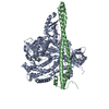
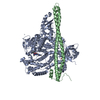
- PDB-1ky4: S-Adenosylhomocysteine hydrolase refined with noncrystallographic... -
+ Open data

- Basic information
| Entry | Database: PDB / ID: 1ky4 | ||||||
|---|---|---|---|---|---|---|---|
| Title | S-Adenosylhomocysteine hydrolase refined with noncrystallographic restraints | ||||||
 Components Components | S-adenosylhomocysteine hydrolase | ||||||
 Keywords Keywords | HYDROLASE / S-adenosylhomocysteine | ||||||
| Function / homology |  Function and homology information Function and homology informationadenosylselenohomocysteinase activity / : / Sulfur amino acid metabolism / S-methylmethionine cycle / circadian sleep/wake cycle / adenyl nucleotide binding / chronic inflammatory response to antigenic stimulus / Methylation / adenosylhomocysteinase / adenosylhomocysteinase activity ...adenosylselenohomocysteinase activity / : / Sulfur amino acid metabolism / S-methylmethionine cycle / circadian sleep/wake cycle / adenyl nucleotide binding / chronic inflammatory response to antigenic stimulus / Methylation / adenosylhomocysteinase / adenosylhomocysteinase activity / L-methionine cycle / one-carbon metabolic process / response to nutrient / NAD binding / melanosome / response to hypoxia / copper ion binding / endoplasmic reticulum / identical protein binding / nucleus / cytosol Similarity search - Function | ||||||
| Biological species |  | ||||||
| Method |  X-RAY DIFFRACTION / MOLECULAR REPLACEMENT / Resolution: 2.8 Å X-RAY DIFFRACTION / MOLECULAR REPLACEMENT / Resolution: 2.8 Å | ||||||
 Authors Authors | Takata, Y. / Takusagawa, F. | ||||||
 Citation Citation | Journal: J.Biol.Chem. / Year: 2002 Title: Catalytic Mechanism of S-adenosylhomocysteine Hydrolase. Site-directed mutagenesis of Asp-130, Lys-185, Asp-189, and Asn-190. Authors: Takata, Y. / Yamada, T. / Huang, Y. / Komoto, J. / Gomi, T. / Ogawa, H. / Fujioka, M. / Takusagawa, F. | ||||||
| History |
|
- Structure visualization
| Structure viewer | Molecule: MolmilJmol/JSmol |
|---|
- Downloads & links
-Download
| PDBx/mmCIF format | 1ky4.cif.gz | 331.5 KB | Display | PDBx/mmCIF format |
|---|---|---|---|---|
| PDB format | pdb1ky4.ent.gz | 267.7 KB | Display | PDB format |
| PDBx/mmJSON format | 1ky4.json.gz | Tree view | PDBx/mmJSON format | |
| Others |  Other downloads Other downloads |
-Validation report
| Arichive directory | https://data.pdbj.org/pub/pdb/validation_reports/ky/1ky4ftp://data.pdbj.org/pub/pdb/validation_reports/ky/1ky4 | HTTPS FTP |
|---|
-Related structure data
-Links
 PDBj
PDBj- Assembly
| Deposited unit | 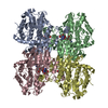
| ||||||||
|---|---|---|---|---|---|---|---|---|---|
| 1 |
| ||||||||
| Unit cell |
| ||||||||
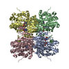
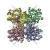
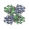
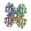
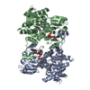
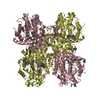
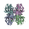
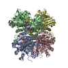
| Details | The biological assembly is a homotetramer. |
-Components
| #1: Protein | Mass: 47465.711 Da / Num. of mol.: 4 Source method: isolated from a genetically manipulated source Source: (gene. exp.)  #2: Chemical | ChemComp-NAD /   Mass: 663.425 Da / Num. of mol.: 4 / Source method: obtained synthetically / Formula: C21H27N7O14P2 / Comment: NAD*YM Mass: 663.425 Da / Num. of mol.: 4 / Source method: obtained synthetically / Formula: C21H27N7O14P2 / Comment: NAD*YM#3: Water | ChemComp-HOH / |  Mass: 18.015 Da / Num. of mol.: 266 / Source method: isolated from a natural source / Formula: H2O Mass: 18.015 Da / Num. of mol.: 266 / Source method: isolated from a natural source / Formula: H2O |
|---|
-Experimental details
-Experiment
| Experiment | Method: X-RAY DIFFRACTION / Number of used crystals: 1 |
|---|
- Sample preparation
| Crystal | Density Matthews: 3.13 Å3/Da / Density % sol: 60.65 % | |||||||||||||||||||||||||||||||||||
|---|---|---|---|---|---|---|---|---|---|---|---|---|---|---|---|---|---|---|---|---|---|---|---|---|---|---|---|---|---|---|---|---|---|---|---|---|
| Crystal grow | Temperature: 297 K / Method: vapor diffusion, hanging drop / pH: 7.2 Details: PEG 6000, pH 7.2, VAPOR DIFFUSION, HANGING DROP, temperature 297K | |||||||||||||||||||||||||||||||||||
| Crystal grow | *PLUS Temperature: 22 ℃Details: used seeding, Komoto, J., (2000) J.Biol.Chem., 275, 32147. | |||||||||||||||||||||||||||||||||||
| Components of the solutions | *PLUS
|
-Data collection
| Diffraction | Mean temperature: 93 K |
|---|---|
| Diffraction source | Source: ROTATING ANODE / Type: RIGAKU RU200 / Wavelength: 1.5418 Å |
| Detector | Type: RIGAKU RAXIS IIC / Detector: IMAGE PLATE / Date: Jan 1, 1999 / Details: Yale mirror |
| Radiation | Protocol: SINGLE WAVELENGTH / Monochromatic (M) / Laue (L): M / Scattering type: x-ray |
| Radiation wavelength | Wavelength: 1.5418 Å / Relative weight: 1 |
| Reflection | Resolution: 2.8→20 Å / Num. all: 58139 / Num. obs: 54797 / % possible obs: 97.5 % / Observed criterion σ(F): 2 / Observed criterion σ(I): 2 / Redundancy: 6.7 % / Rmerge(I) obs: 0.071 / Rsym value: 0.071 / Net I/σ(I): 11.4 |
| Reflection shell | Resolution: 2.8→2.92 Å / Redundancy: 3.1 % / Rmerge(I) obs: 0.16 / Mean I/σ(I) obs: 5 / Num. unique all: 5050 / Rsym value: 0.16 / % possible all: 87 |
| Reflection | *PLUS Rmerge(I) obs: 0.071 |
| Reflection shell | *PLUS Rmerge(I) obs: 0.16 |
- Processing
| Software |
| |||||||||||||||||||||
|---|---|---|---|---|---|---|---|---|---|---|---|---|---|---|---|---|---|---|---|---|---|---|
| Refinement | Method to determine structure: MOLECULAR REPLACEMENT / Resolution: 2.8→8 Å / σ(F): 2 / Stereochemistry target values: Engh & Huber
| |||||||||||||||||||||
| Refine analyze | Luzzati coordinate error obs: 0.04 Å | |||||||||||||||||||||
| Refinement step | Cycle: LAST / Resolution: 2.8→8 Å
| |||||||||||||||||||||
| Refine LS restraints |
| |||||||||||||||||||||
| LS refinement shell | Refine-ID: X-RAY DIFFRACTION
| |||||||||||||||||||||
| Refinement | *PLUS Num. reflection Rfree: 4950 / Rfactor all: 0.23 / Rfactor obs: 0.229 / Rfactor Rfree: 0.278 / Rfactor Rwork: 0.228 | |||||||||||||||||||||
| Solvent computation | *PLUS | |||||||||||||||||||||
| Displacement parameters | *PLUS | |||||||||||||||||||||
| Refine LS restraints | *PLUS
| |||||||||||||||||||||
| LS refinement shell | *PLUS Rfactor Rfree: 0.3353 / Rfactor Rwork: 0.2821 |
