 Movie
Movie Controller

[English] 日本語
 Yorodumi
- PDB-1k0u: Inhibition of S-adenosylhomocysteine Hydrolase by "acyclic sugar"... -
+ Open data

- Basic information
| Entry | Database: PDB / ID: 1k0u | |||||||||
|---|---|---|---|---|---|---|---|---|---|---|
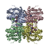
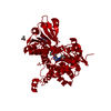
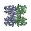
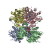
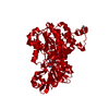
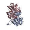
| Title | Inhibition of S-adenosylhomocysteine Hydrolase by "acyclic sugar" Adenosine Analogue D-eritadenine | |||||||||
 Components Components | S-ADENOSYL-L-HOMOCYSTEINE HYDROLASE | |||||||||
 Keywords Keywords | HYDROLASE / S-adenosylhomocysteine / D-eritadenine / inhibitor | |||||||||
| Function / homology |  Function and homology information Function and homology informationadenosylselenohomocysteinase activity / : / Sulfur amino acid metabolism / S-methylmethionine cycle / circadian sleep/wake cycle / adenyl nucleotide binding / chronic inflammatory response to antigenic stimulus / Methylation / adenosylhomocysteinase / adenosylhomocysteinase activity ...adenosylselenohomocysteinase activity / : / Sulfur amino acid metabolism / S-methylmethionine cycle / circadian sleep/wake cycle / adenyl nucleotide binding / chronic inflammatory response to antigenic stimulus / Methylation / adenosylhomocysteinase / adenosylhomocysteinase activity / L-methionine cycle / one-carbon metabolic process / response to nutrient / NAD binding / melanosome / response to hypoxia / copper ion binding / endoplasmic reticulum / identical protein binding / nucleus / cytosol Similarity search - Function | |||||||||
| Biological species |  | |||||||||
| Method |  X-RAY DIFFRACTION / MOLECULAR REPLACEMENT / Resolution: 3 Å X-RAY DIFFRACTION / MOLECULAR REPLACEMENT / Resolution: 3 Å | |||||||||
 Authors Authors | Takusagawa, F. / Huang, Y. / Komoto, J. / Takata, Y. / Gomi, T. / Ogawa, H. / Fujioka, M. / Powell, D. | |||||||||
 Citation Citation | Journal: J.Biol.Chem. / Year: 2002 Title: Inhibition of S-adenosylhomocysteine hydrolase by acyclic sugar adenosine analogue D-eritadenine. Crystal structure of S-adenosylhomocysteine hydrolase complexed with D-eritadenine. Authors: Huang, Y. / Komoto, J. / Takata, Y. / Powell, D.R. / Gomi, T. / Ogawa, H. / Fujioka, M. / Takusagawa, F. | |||||||||
| History |
|
- Structure visualization
| Structure viewer | Molecule: MolmilJmol/JSmol |
|---|
- Downloads & links
-Download
| PDBx/mmCIF format | 1k0u.cif.gz | 630.3 KB | Display | PDBx/mmCIF format |
|---|---|---|---|---|
| PDB format | pdb1k0u.ent.gz | 517.3 KB | Display | PDB format |
| PDBx/mmJSON format | 1k0u.json.gz | Tree view | PDBx/mmJSON format | |
| Others |  Other downloads Other downloads |
-Validation report
| Arichive directory | https://data.pdbj.org/pub/pdb/validation_reports/k0/1k0uftp://data.pdbj.org/pub/pdb/validation_reports/k0/1k0u | HTTPS FTP |
|---|
-Related structure data
| Related structure data | 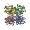 1d4fS S: Starting model for refinement |
|---|---|
| Similar structure data |
-Links
 PDBj
PDBj- Assembly
| Deposited unit | 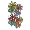
| ||||||||
|---|---|---|---|---|---|---|---|---|---|
| 1 | 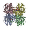
| ||||||||
| 2 | 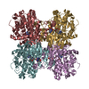
| ||||||||
| Unit cell |
| ||||||||
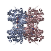
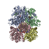
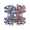
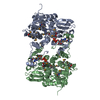
| Details | The asymmetric unit contains two tetrameric enzymes, i.e., contains eight identical subunits. |
-Components
| #1: Protein | Mass: 47465.711 Da / Num. of mol.: 8 Source method: isolated from a genetically manipulated source Source: (gene. exp.)  #2: Chemical | ChemComp-NAD /   Mass: 663.425 Da / Num. of mol.: 8 / Source method: obtained synthetically / Formula: C21H27N7O14P2 / Comment: NAD*YM Mass: 663.425 Da / Num. of mol.: 8 / Source method: obtained synthetically / Formula: C21H27N7O14P2 / Comment: NAD*YM#3: Chemical | ChemComp-DEA /   Mass: 253.215 Da / Num. of mol.: 8 / Source method: obtained synthetically / Formula: C9H11N5O4 / Comment: inhibitor*YM Mass: 253.215 Da / Num. of mol.: 8 / Source method: obtained synthetically / Formula: C9H11N5O4 / Comment: inhibitor*YM#4: Water | ChemComp-HOH / |  Mass: 18.015 Da / Num. of mol.: 474 / Source method: isolated from a natural source / Formula: H2O Mass: 18.015 Da / Num. of mol.: 474 / Source method: isolated from a natural source / Formula: H2O |
|---|
-Experimental details
-Experiment
| Experiment | Method: X-RAY DIFFRACTION / Number of used crystals: 1 |
|---|
- Sample preparation
| Crystal | Density Matthews: 2.24 Å3/Da / Density % sol: 45.17 % | ||||||||||||||||||||||||||||||
|---|---|---|---|---|---|---|---|---|---|---|---|---|---|---|---|---|---|---|---|---|---|---|---|---|---|---|---|---|---|---|---|
| Crystal grow | Temperature: 295 K / Method: vapor diffusion, hanging drop / pH: 7.2 Details: PEG 4000, pH 7.2, VAPOR DIFFUSION, HANGING DROP, temperature 295K | ||||||||||||||||||||||||||||||
| Crystal grow | *PLUS Temperature: 22 ℃ | ||||||||||||||||||||||||||||||
| Components of the solutions | *PLUS
|
-Data collection
| Diffraction | Mean temperature: 93 K |
|---|---|
| Diffraction source | Source: ROTATING ANODE / Type: RIGAKU RU200 / Wavelength: 1.5418 Å |
| Detector | Type: RIGAKU RAXIS IIC / Detector: IMAGE PLATE / Date: Aug 20, 1999 / Details: mirrors |
| Radiation | Monochromator: YALE MIRRORS / Protocol: SINGLE WAVELENGTH / Monochromatic (M) / Laue (L): M / Scattering type: x-ray |
| Radiation wavelength | Wavelength: 1.5418 Å / Relative weight: 1 |
| Reflection | Resolution: 3→10 Å / Num. all: 59076 / Num. obs: 56933 / % possible obs: 85 % / Observed criterion σ(F): 3 / Observed criterion σ(I): 3 / Redundancy: 2.1 % / Biso Wilson estimate: 10 Å2 / Rmerge(I) obs: 0.085 / Rsym value: 0.085 / Net I/σ(I): 6.2 |
| Reflection shell | Resolution: 3→3.1 Å / Redundancy: 1.8 % / Rmerge(I) obs: 0.19 / Mean I/σ(I) obs: 3.1 / Rsym value: 0.19 / % possible all: 82 |
| Reflection | *PLUS Lowest resolution: 10 Å / Num. obs: 59048 / % possible obs: 91.5 % / Num. measured all: 241990 |
- Processing
| Software |
| |||||||||||||||||||||||||
|---|---|---|---|---|---|---|---|---|---|---|---|---|---|---|---|---|---|---|---|---|---|---|---|---|---|---|
| Refinement | Method to determine structure: MOLECULAR REPLACEMENT Starting model: 1D4F Resolution: 3→10 Å / Isotropic thermal model: Isotropic / Cross valid method: THROUGHOUT / σ(F): 2 / Stereochemistry target values: Engh & Huber
| |||||||||||||||||||||||||
| Displacement parameters | Biso mean: 5.3 Å2
| |||||||||||||||||||||||||
| Refine analyze | Luzzati sigma a obs: 0.34 Å | |||||||||||||||||||||||||
| Refinement step | Cycle: LAST / Resolution: 3→10 Å
| |||||||||||||||||||||||||
| Refine LS restraints |
| |||||||||||||||||||||||||
| LS refinement shell | Resolution: 3→10 Å / Rfactor Rfree error: 0.01
| |||||||||||||||||||||||||
| Software | *PLUS Name: X-PLOR / Version: 3.851 / Classification: refinement | |||||||||||||||||||||||||
| Refinement | *PLUS Highest resolution: 3 Å / Lowest resolution: 10 Å / σ(F): 2 | |||||||||||||||||||||||||
| Solvent computation | *PLUS | |||||||||||||||||||||||||
| Displacement parameters | *PLUS Biso mean: 5.3 Å2 | |||||||||||||||||||||||||
| Refine LS restraints | *PLUS
| |||||||||||||||||||||||||
| LS refinement shell | *PLUS Highest resolution: 3 Å / Lowest resolution: 10 Å / Rfactor Rfree: 0.265 / Rfactor Rwork: 0.208 |
